Description
Developmental Juvenile Osteology (2nd Ed.)
Authors: Cunningham Craig, Scheuer Louise, Black Sue
Language: English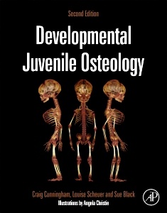
Subjects for Developmental Juvenile Osteology:
Keywords
Juvenile; Osteology; Development; Skeleton; Age estimation; Identification; Ossification; Epiphyseal fusion; Growth; Sub-adult
173.83 €
In Print (Delivery period: 14 days).
Add to cart the book of Cunningham Craig, Scheuer Louise, Black Sue
630 p. · 21.5x27.6 cm · Hardback
Description
/li>Contents
/li>Readership
/li>Biography
/li>Comment
/li>
Developmental Juvenile Osteology was created as a core reference text to document the development of the entire human skeleton from early embryonic life to adulthood.
In the period since its first publication there has been a resurgence of interest in the developing skeleton, and the second edition of Developmental Juvenile Osteology incorporates much of the key literature that has been published in the intervening time.
The main core of the text persists by describing each individual component of the human skeleton from its embryological origin through to its final adult form. This systematic approach has been shown to assist the processes of both identification and age estimation and acts as a core source for the basic understanding of normal human skeletal development. In addition to this core, new sections have been added where there have been significant advances in the field.
2 Skeletal Development and Aging
3. Bone Development
4. Early Embryological Development
5. The Skull
6. Dentition
7. The Vertebral Column
8. The Thorax
9. The Pectoral Girdle
10. The Upper Limb
11. The Pelvic Girdle
12. The Lower Limb
Professor Louise Scheuer is a retired anatomist and forensic anthropologist who taught at several London medical schools including 20 years at St.Thomas’s Hospital Medical School and the Medical School of University College London. She is a past President of the British Association of Clinical Anatomists and holds an Honorary (Chair) Professorship at Dundee University.
She is a member of the Anatomical Society of Great Britain and Northern Ireland, the British Association of Clinical Anatomy and a Fellow of the Royal Anthropological Institute and of the Royal Society of Medicine.
She and Sue (Prof.) Black held a Leverhulme Grant for the conservation and re-evaluation of the St. Bride’s Church skeletal collection.
She has worked with forensic pathologists, coroners and police on the identification of human remains and was a forensic anthropologist in the British Foreign and Com
- Identifies every component of the juvenile skeleton, by providing a detailed analysis of development and ageing and a detailed description of each bone in four ways: adult bone, early development, ossification and practical notes
- New chapters and updated sections covering the dentition, age estimation in the living and bone histology
- An updated bibliography documenting the research literature that has contributed to the field over the past 15 years since the publication of the first edition
- Heavily illustrated, including new additions