Description
Breast Imaging Case Series: Radiologic & Pathologic Correlations
Authors: Joshi Surekha, Kulkarni Kirti
Language: English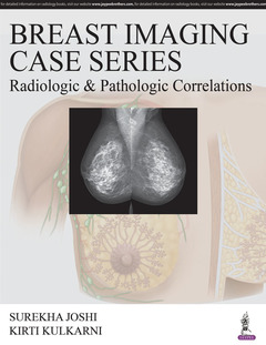
Subjects for Breast Imaging Case Series: Radiologic & Pathologic...:
Publication date: 05-2019
350 p. · 21.5x27.9 cm · Hardback
Publication Abandoned
350 p. · 21.5x27.9 cm · Hardback
Publication Abandoned
Description
/li>Contents
/li>Biography
/li>
This book is a comprehensive guide to breast imaging for clinicians and
trainees. Presented as a series of cases, the text follows a unique
approach by correlating radiological pathological findings. Beginning with
an overview of breast screening, the book covers all imaging techniques
mammography, ultrasound, MRI, and new technologies including digital
breast tomosynthesis and molecular imaging.
The following sections explain different types of breast cancer, staging, surgical planning, unusual lesions, and imaging in pregnancy and lactation, and with breast implants. A chapter is dedicated to evaluation of the male breast.
Authored by recognised specialists from Tennessee and Chicago, the book features numerous radiological images to assist understanding. Each case is different, creating a unique radiology-pathology correlation pattern.
- 1. Screening Of Breast Cancer
- 2. Diagnostic Mammogram And Ultrasound
- 3. Image Guided Intervention
- 4. Establishing Concordance After Image Guided Biopsy
- 5. High Risk Lesions: ADH, ALH, Papillary Lesions, Radial Scar
- 6. Imaging Features Of Breast Cancers
- 7. Staging Of Breast Cancer
- 8. Surgical Planning, Sentinel Node Injection
- 9. Imaging Surveillance After Breast Cancer Diagnosis
- 10. Breast Imaging During Pregnancy And Lactation
- 11. Imaging Of Breast Implants And The Altered Breast
- 12. Evaluation Of The Male Breast
- 13. Unusual Breast Lesions: Pagets, Lymphoma, Sarcoma, Metastasis, Phylloides, Granulomatous Mastitis, Mondors Disease
- 14. Artifacts: Mammography, Ultrasound And MRI
- 15. Special Considerations: PEM, BSGI ( Molecular Imaging) And Digital Contrast Enhanced Mammography
Surekha Joshi MD
Breast Imaging & Intervention, Diagnostic Imaging, Assistant Professor, University of Tennessee Health Science Centre, USA
Kirti Kulkarni MD
Assistant Professor, Section of Abdominal and Breast Imaging, Director, Breast Imaging Fellowship Program, Department of Radiology, University of Chicago Medicine, Chicago, USA
© 2024 LAVOISIER S.A.S.
These books may interest you
Breast Imaging 241.19 €