


Description
Color Atlas of Dermoscopy
Author: Cabo Horacio
Language: English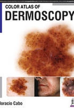
Subject for Color Atlas of Dermoscopy:
Approximative price 206.02 €
In Print (Delivery period: 14 days).
Add to cart the book of Cabo Horacio
400 p. · 21.5x27.9 cm · Hardback
Description
/li>Contents
/li>Readership
/li>Biography
/li>
Dermoscopy is a non-invasive, widely used diagnostic tool that aids the diagnosis of skin lesions and is proven to increase the accuracy of melanoma diagnosis.
This colour atlas is a comprehensive guide to the diagnosis of skin lesions and melanomas using a dermoscope.
Beginning with an introduction to the use of the dermascope, the following chapters teach clinicians how to recognise dermoscopic criteria, colours and patterns, how to diagnose different types of lesions and calculate diagnostic algorithms.
The finals sections cover related topics including entomodermatoscopy, inflammatoscopy, trichoscopy and capilaroscopy.
This highly useful resource is enhanced by more than 1000 clinical images and illustrations.
Key points
- Comprehensive guide to diagnosis of skin lesions and melanomas using a dermoscope
- Teaches clinicians how to recognise dermoscopic criteria
- Covers related dermatoscopic topics
- Includes more than 1000 images and illustrations
- Why Use The Dermatoscope
- Structures, Patterns, Criteria And Colours
- Vascular Patterns
- Dermoscopy: A Two-Step Procedure
- Non Melanocytic Lesions
- Melanocytic Lesions
- Melanoma Simulators
- Combined Lesions
- Special Locations
- Diagnostic Algorithms
- Dermoscopy Follow-Up And Total Body Photography
- Revised Pattern Analysis
- Entomodermatoscopy
- Inflammatoscopy
- Tricoscopy
- Capilaroscopy
- Reflectance Confocal Microscopy In Vivo
- Chaos & Clues
Horacio Cabo MD
Professor of Dermatology, University of Buenos Aires; Dermatology Specialist, Buenos Aires, Argentina
These books may interest you
Atlas of Dermoscopy 135.14 €
Pocket Guide to Dermoscopy 47.10 €
DermoscopyText and Atlas 185.59 €