Description
Diagnostic Ultrasound, 2-Volume Set (6th Ed.)
Coordinators: Rumack Carol M., Levine Deborah
Language: English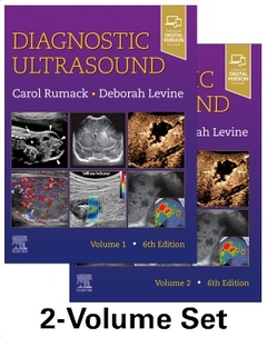
Subjects for Diagnostic Ultrasound, 2-Volume Set:
Keywords
<; P>; Ultrasound; Doppler; Liver; Spleen; Gallbladder; Pancreas; Kidney; Thyroid Gland; Breast; Musculoskeletal; Obstetric Imaging; Fetal Imaging; Doppler Sonography; Neonatal<; /P>

2280 p. · 21.5x27.6 cm · Hardback
Description
/li>Contents
/li>Biography
/li>
Covers all aspects of diagnostic ultrasound with sections for Physics; Abdominal, Pelvic, Small Parts, Vascular, Obstetric, and Pediatric Sonography.
Contains 5,000 images throughout, including 2D and 3D imaging as well as the use of contrast agents and elastography.
Includes a new section on setting up a contrast lab for clinical practice and a new chapter on hemodialysis.
Features new coverage of the parotid, salivary, and submandibular glands, as well as the retroperitoneum, which now includes a section on endoleaks with ultrasound contrast.
Uses a straightforward writing style and extensive image panels with correlative findings.
Includes 400 video clips showing real-time scanning of anatomy and pathology.
An eBook version is included with purchase. The eBook allows you to access all of the text, figures and references, with the ability to search, customize your content, make notes and highlights, and have content read aloud.
All chapters will have updated images
I. PHYSICS 1. Physics of Ultrasound (*Heavily revised) 2. Biologic Effects and Safety (*Heavily revised) 3. Ultrasound Contrast Agents and Setting Up Your Contrast Practice (includes new section on setting up a contrast lab for clinical practice) II. ABDOMINAL, PELVIC, AND THORACIC SONOGRAPHY 4. The Liver 5. The Spleen 6. The Biliary Tree and Gallbladder 7. The Pancreas 8. The Gastrointestinal Tract 9. The Kidney and Urinary Tract 10. The Prostrate 11. The Adrenal Glands 12. The Retroperitoneum (will include a section on endoleaks with US contrast) 13. Dynamic Ultrasound of Hernias of the Groin and Anterior Abdominal Wall 14. The Peritoneum 15. The Uterus 16. The Adnexa 17. Ultrasound-Guided Biopsy of Chest Abdomen and Pelvis 18. Organ Transplantation III. SMALL PARTS, CAROTID ARTERY, AND PERIPHERAL VESSEL SONOGRAPHY 19. The Thyroid Gland 20. Other glands in the neck (which was just parathyroid now includes other glands in the neck (parotid, salivary and submandibular glands) 21. The Breast 22. The Scrotum 23. Overview of Musculoskeletal Ultrasound Techniques and Applications 24. The Shoulder 25. Musculoskeletal Interventions 26. The Extracranial Cerebral Vessels 27. The Peripheral Arteries (*Heavily revised) 28. The peripheral veins 29. Hemodialysis (new Chapter)
IV. OBSTETRIC AND FETAL SONOGRAPHY 30. Overview of Obstetric Imaging 31. The First Trimester 32. Chromosomal Abnormalities 33. Multifetal Pregnancy 34. The Fetal Face and Neck 35. The Fetal Brain 36. The Fetal Spine 37.The Fetal Chest 38. The Fetal Heart 39. The Fetal Abdominal Wall and Gastrointestinal Tract 40. The Fetal Urogenital Tract 41. The Fetal Musculoskeletal System 42. Fetal Hydrops 43. Fetal Measurements: Normal and Abnormal Fetal Growth and Assessment of Fetal Well-Being 44. Sonographic Evaluation of the Placenta 45. Cervical Ultrasound and Preterm Birth V. PEDIATRIC SONOGRAPHY 46. Neonatal and Infant Brain Imaging 47. Doppler Sonography of the Neonatal and Pediatric Brain 48. The Pediatric Head and Neck 50. The Pediatric Spinal Canal 51 The Pediatric Chest 52. The Pediatric Liver and Spleen 53. The Pediatric Kidney and Adrenal Glands 54.The Pediatric Gastrointestinal Tract 55. Pediatric Pelvic Sonography 56.The Pediatric Hip and Musculoskeletal Ultrasound 57. Pediatric Interventional Sonograph
Deborah Levine, MD, is Vice Chair of Academic Affairs in the Department of Radiology at Beth Israel Deaconess Medical Center and Professor of Radiology at Harvard Medical School in Boston. Dr Levine's clinical experience is in obstetric and gynecologic ultrasound, and her research is focused on fetal MRI imaging. She is an American College of Radiology Chancellor, Chair of the American College of Radiology Commission on Ultrasound, and a fellow of the American Institute of Ultrasound in Medicine and the Society of Radioloigists in Ultrasound.
These books may interest you
Diagnostic Ultrasound: Head and Neck 261.92 €