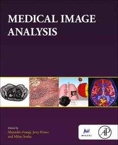
Description
Medical Image Analysis
The MICCAI Society book Series
Coordinators: Frangi Alejandro, Prince Jerry, Sonka Milan
Language: English
Subjects for Medical Image Analysis:
Publication date: 09-2023
698 p. · 19x23.4 cm · Paperback
698 p. · 19x23.4 cm · Paperback
Description
/li>Contents
/li>Readership
/li>Biography
/li>Comment
/li>
Medical imaging is increasingly at the base of many breakthroughs in biomedical sciences, becoming a fundamental enabling technology of biomedical scientific progress. Medical Image Analysis presents practical knowledge on medical image computing and analysis and is written by top educators and experts in the field. This text is a modern, practical, broad, and self-contained reference that conveys a mix of essential methodological concepts within different medical domains, reflecting the nature of the discipline today, making it suitable as a course text and a self-learning resource.
PART I Introductory topics
1. Medical imaging modalities
2. Mathematical preliminaries
3. Regression and classification
4. Estimation and inference
PART II Image representation and processing
5. Image representation and 2D signal processing
6. Image filtering: enhancement and restoration
7. Multiscale and multiresolution analysis
PART III Medical image segmentation
8. Statistical shape models
9. Segmentation by deformable models
10. Graph cut-based segmentation
PART IV Medical image registration
11. Points and surface registration
12. Graph matching and registration
13. Parametric volumetric registration
14. Non-parametric volumetric registration
15. Image mosaicking
PART V Machine learning in medical image analysis
16. Deep learning fundamentals
17. Deep learning for vision and representation learning
18. Deep learning medical image segmentation
19. Machine learning in image registration
PART VI Advanced topics in medical image analysis
20. Motion and deformation recovery and analysis
21. Imaging Genetics
PART VII Large-scale databases
22. Detection and quantitative enumeration of objects from large images
23. Image retrieval in big image data
PART VIII Evaluation in medical image analysis
24. Assessment of image computing methods
Researchers in Medical Image computing and analysis; Ideal Text-Ref for courses on medical image analysis in BEng/MEng/MSc programs in Biomedical Engineering, Medical Physics, Medical Engineering, or Computational Bioengineering
Alejandro F. Frangi is the Bicentennial Turing Chair in Computational Medicine and Royal Academy of Engineering Chair in Emerging Technologies at The University of Manchester, Manchester, UK, with joint appointments at the Schools of Engineering (Department of Computer Science), Faculty of Science and Engineering, and the School of Health Sciences (Division of Informatics, Imaging and Data Science), Faculty of Biology, Medicine and Health.
He is a Turing Fellow of the Alan Turing
Institute. He holds an Honorary Chair at KU Leuven in the Departments of Electrical Engineering (ESAT) and Cardiovascular Science. He is IEEE Fellow (2014), EAMBES Fellow (2015), SPIE Fellow (2020), MICCAI Fellow (2021), and Royal Academy of Engineering Fellow (2023). The IEEE Engineering in Medicine and Biology Society awarded him the Early Career Award (2006) and Technical Achievement Award (2021). Professor Frangi’s primary research interests are in medical image analysis and modeling, emphasising machine learning (phenomenological models) and computational physiology (mechanistic models). He is an expert in statistical
shape modeling, computational anatomy, and image-based computational physiology, delivering novel insights and impact across various imaging modalities and diseases,
particularly on cardiovascular MRI, cerebrovascular MRI/CT/3DRA, and musculoskeletal CT/DXA. He is a co-founder of adsilico Ltd., and his work led to products
commercialized by GalgoMedical SA. He has published over 285 peer-reviewed papers in scientific journals with over 34,000 citations and has an h-index of 75.
Jerry L. Prince is the William B. Kouwenhoven Professor in the Department of Electrical and Computer Engineering at Johns Hopkins University. He is Director of the Image Analysis and Communications Laboratory (IACL). He also holds joint appointments in the Departments of Radiology and Radiological Science, Biomedical Engineering, Computer Scienceand Applied Mathematics and S
- An authoritative presentation of key concepts and methods from experts in the field
- Sections clearly explaining key methodological principles within relevant medical applications
- Self-contained chapters enable the text to be used on courses with differing structures
- A representative selection of modern topics and techniques in medical image computing
- Focus on medical image computing as an enabling technology to tackle unmet clinical needs
- Presentation of traditional and machine learning approaches to medical image computing
© 2024 LAVOISIER S.A.S.


