Description
Spectral Domain Optical Coherence Tomography in Macular Diseases, 1st ed. 2016
Coordinators: Meyer Carsten H., Saxena Sandeep, Sadda SriniVas R
Language: English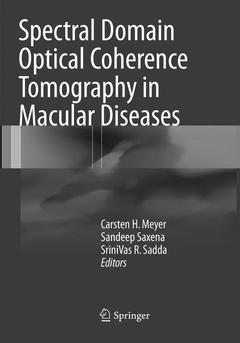
Subject for Spectral Domain Optical Coherence Tomography in Macular...:

Publication date: 07-2018
Support: Print on demand
Publication date: 12-2016
443 p. · 17.8x25.4 cm · Hardback
Description
/li>Contents
/li>Biography
/li>Comment
/li>
This book aims to build concepts and create a solid foundation in the field of optical coherence tomography (OCT) for the general ophthalmologists as well as for the resident trainees and fellows. The chapters are written by leading international authorities in a style comprehensible to a broad audience. Numerous clinical pictures and SD-OCT scans help elucidate various clinical entities.OCT is the optical analog of ultrasound imaging and has emerged as a powerful imaging technique that enables non-invasive, in-vivo, high-resolution, cross-sectional imaging in retinal tissue. A new generation spectral domain optical coherence tomography (SD-OCT) technology has now been developed, representing a quantum leap in resolution and speed, achieving in vivo optical biopsy. i.e. the visualization of tissue architectural morphology in situ and in real time. This book encompasses the role of SD-OCT in both medical and surgical macular disorders.
The book is meant coherent and comprehensive for both vitreoretinal specialists as well as general ophthalmologists.
SriniVas R. Sadda, MD, is the President and Chief Scientific Officer of the Doheny Eye Institute, the Stephen J. Ryan – Arnold and Mabel Beckman Endowed Chair, and Professor of Ophthalmology at the University of California – Los Angeles (UCLA) Geffen School of Medicine. He has more than 280 peer-reviewed publications and 13 book chapters. Dr. Sadda also serves as an editorial board member of Ophthalmic Surgery, Lasers & Imaging, Retina, and Ophthalmology. He is also an editor of the 5th edition of the Ryan’s Retina textbook.
Provides point-to-point correlation of macular diseases and SD-OCT
Covers 3-dimentional imaging and segmentation analysis
Provides planning of surgery using SD-OCT