Description
7.0 Tesla MRI Brain Atlas (2nd Ed., 2nd ed. 2015)
In-vivo Atlas with Cryomacrotome Correlation
Coordinator: Cho Zang-Hee
Language: English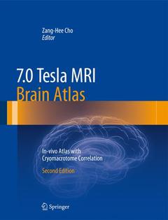
Subjects for 7.0 Tesla MRI Brain Atlas:

Publication date: 01-2015
544 p. · 22.7x30 cm · Hardback
544 p. · 22.7x30 cm · Hardback
Description
/li>Contents
/li>Biography
/li>Comment
/li>
The inaugural publication of the 7.0 Tesla MRI Brain Atlas: In Vivo Atlas with Cryomacrotome Correlation in 2010 provided readers with a spectacular source of ultra-high resolution images revealing a wealth of details of the brainstem and midbrain structures. This second edition contributes additional knowledge gained as a result of technologic advances and recent research. To facilitate identification and comparison of brain structures and anatomy, a detailed coordination matrix is featured in each image. Updated axial, sagittal, and coronal images are also included. This state-of-the-art and user-friendly reference will provide researchers and clinicians with important new perspectives.
Foreword.- Preface.- Preface of the 2nd Edition.- Introduction.- 1. Reference of Brain Image Setting.- 2. Orientation of Brain Images.- I. Orientation of the image sections and planes.- II. Standard of the sectional brain image planes and sizes.- III. Adjustment of MRI brain to the reference brain.- IV. Terminology & Labeling.- V. Data collection system for clinicopathologic brain mapping.- VI. Figures (Figs. 1, 2, 3).- 3. Sources of Brain Images.- I. In vivo Images using 7.0T MRI.- II. Cadaver Images by Cryomacrotome.- III. Image reconstruction and volume rendering.- 4. 3D Images by Volume Rendering.- I. Coronal, Sagittal and Axial Cuts- Cadaver (Fig. 4).- II. Coronal, Sagittal and Axial Cuts- MRI (Fig. 5).- III. Sulcus and Gyrus- MRI (Fig. 6).- IV.Brodmann areas-MRI (Fig. 7).- Acknowledgements.- Chapter I. Coronal Images of Cadaver & Human Brain of 7.0T MRI In Vivo.- Chapter II. Sagittal Images of Cadaver & Human Brain of 7.0T MRI In Vivo.- Chapter III. Axial Images of Cadaver & Human Brain of 7.0T MRI In Vivo.- References.- Abbreviations.- Index.
Zang-Hee Cho, Ph.D, is a Korean neuroscientist who developed the first Ring-PET scanner and the scintillation detector BGO. More recently, Cho developed the first PET-MRI fusion molecular imaging device for neuro-molecular imaging. He has held several academic positions in Sweden, the USA and South Korea.
Revised edition with updated in vivo images
Features a coordination matrix in each image, facilitating identification of brain structures and anatomy
User-friendly format and size
© 2024 LAVOISIER S.A.S.