


Description
7.0 Tesla MRI Brain White Matter Atlas (2nd Ed., 2nd ed. 2015)
Language: English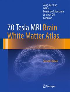
Subjects for 7.0 Tesla MRI Brain White Matter Atlas:

Publication date: 01-2015
457 p. · 22.7x30 cm · Hardback
457 p. · 22.7x30 cm · Hardback
Description
/li>Contents
/li>Biography
/li>Comment
/li>
The introduction of techniques that permit visualization of the human nervous system is one of the foremost advances in neuroscience and brain-related research. Among the most recent significant developments in this respect are ultra-high field MRI and the image post-processing technique known as track density imaging (TDI). It is these techniques (including super-resolution TDI) which represent the two major components of 7.0 Tesla MRI ? Brain White Matter Atlas. This second edition of the atlas has been revised and updated to fully reflect current application of these technological advancements in order to visualize the nervous system and the brain with the finest resolution and sensitivity. Exquisitely detailed color images offer neuroscientists, neurologists, and neurosurgeons a superb resource that will be of value both for the purpose of research and for the treatment of common brain diseases such as Alzheimer's disease and multiple sclerosis.
Introduction.- 1. MR Diffusion Tensor Imaging (DTI) and Track-Density Imaging (TDI).- a. MR Diffusion Tensor Imaging (DTI) and Super-Resolution Track-Density Imaging (TDI).- b. Track-Density Imaging (TDI) – Examples of DTI and TDI-1.- 2. Views, Directions, and Orientations of Brain Images.- a. Views and Directions of the Brain Image.- b. Definition of the Central Intercommissural Line.- c. Volume Rendered 3D Images.- Acknowledgments.- PART 1. Coronal Images of Tractography and Corresponding In-Vivo 7.0-T MRI Anatomy.- PART 2. Sagittal Images of Tractography and Corresponding In-Vivo 7.0-T MRI Anatomy.- PART 3. Axial Images of Tractography and Corresponding In-Vivo 7.0-T MRI Anatomy.
Zang-Hee Cho, Ph.D, is a Korean neuroscientist who developed the first Ring-PET scanner and the scintillation detector BGO. More recently, Cho developed the first PET-MRI fusion molecular imaging device for neuro-molecular imaging. He has held several academic positions in Sweden, the USA and South Korea. He published the 7.0 Tesla MRI Brain Atlas with Springer in 2010 (currently out of stock).
Depicts the visualization of brain white matter with the latest 7.0 T MRI and TDI techniques Represents a useful addition to brain research and clinical settings, such as the Human Connectome Project Contains a wealth of exquisitely detailed color images
© 2024 LAVOISIER S.A.S.
These books may interest you
Atlas of Functional Neuroanatomy 87.11 €
Neuroanatomy text and atlas 69.87 €