Description
Color Atlas of Veterinary Histology (3rd Ed.)
Authors: Bacha Jr. William J., Bacha Linda M.
Language: English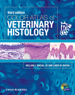
Subject for Color Atlas of Veterinary Histology:
Keywords
students; foundation; cytologic; histologic; provide; preparations; practical; histology; reference; domestic; normal; color atlas; veterinary; common; benchside; species; eight; images; edition; third; new; throughout; types; tissues
Approximative price 109.84 €
In Print (Delivery period: 14 days).
Add to cart the book + cd of Bacha Jr. William J., Bacha Linda M.
368 p. · 22.1x27.7 cm · Hardback
Description
/li>Contents
/li>Readership
/li>Biography
/li>
For the first time, the more than 900 photomicrographs are available digitally in an interactive atlas on CD, offering images available for download with zoom capability. The new edition of this veterinary-specific histology atlas provides veterinary and veterinary technician students with an essential pictorial resource for interpreting histologic preparations.
2 Epithelium
3 Connective Tissue Proper and Embryonic Connective Tissue
4 Cartilage
5 Bone Tissue
6 Blood
7 Bone Marrow
8 Muscle Tissue
9 Nervous System
10 Cardiovascular System
11 Lymphatic System
12 Integument
13 Digestive System
14 Urinary System
15 Respiratory System
16 Endocrine System
17 Male Reproductive System
18 Female Reproductive System
19 The Eye
20 The Ear
Glossary
Bibliography
Index
Linda M. Bacha, VMD, is Assistant Professor I of Biology in the Department of Biology, Camden County College, Blackwood, New Jersey.
These books may interest you
Color Atlas of Equine Pathology 164.63 €
Invertebrate Histology 220.32 €