Description
Forensic Microscopy for Skeletal Tissues, Softcover reprint of the original 1st ed. 2012
Methods and Protocols
Methods in Molecular Biology Series, Vol. 915
Coordinator: Bell Lynne S.
Language: English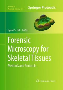
Subject for Forensic Microscopy for Skeletal Tissues:

Support: Print on demand
269 p. · 17.8x25.4 cm · Paperback
Description
/li>Contents
/li>Comment
/li>
Forensic anthropology deals with human remains usually in the skeletonized form. The application of microscopy to skeletal tissues is well established and used routinely in biomedical science. Its adaptation to forensic questions is an increasing area of interest, and publications utilizing microscopy have increased in the scientific literature. In Forensic Microscopy for Skeletal Tissues: Methods and Protocols, expert researchers in the forensic, archeological and paleontological disciplines, and detail many of the methods which are now commonly used to study skeletal material. These methods include differing forms of light, confocal, scanning electron and transmission electron microscopy. Written in the highly successful Methods in Molecular Biology? series format, chapters include introductions to their respective topics, lists of the necessary materials and reagents, step-by-step, readily reproducible laboratory protocols, and key tips on troubleshooting and avoiding known pitfalls.
Authoritative and practical, Forensic Microscopy for Skeletal Tissues: Methods and Protocols bring together differing forms of microscopy that are used in association with forensic anthropology, or have relevance to questions concerning forensic anthropology.
Scanning Electron Microscopy - Preparation and Imaging for SEM.-Plastic Embedding and Polishing of Bone for Reflected Light and Electron Microscopy.-Preparation of Mineralised Tissue for Light Microscopy.-Bone Pathology
Kim Piper and Gail Valentine.-A Histological Method That Can Be Used to Estimate the Time Taken to Form the Crown of a Permanent Tooth.-The Determination of Age in Subadult from the Rib Cortical Microstructure.-Rib Histomorphometry for Adult Age Estimation.-Age Estimation using Tooth Cementum Annulation.-Dental Topographic Analysis of the Molar Teeth of Primates.-Chemical Analyses of Fossil Bone.-Identifying Post Mortem Microstructural Change to Skeletal and Dental Tissues using Backscattered Electron Imaging.-How to Approach Peri-Mortem Injury and Other Modification.-Light Microscopy of Microfractures in Burned Bone.-A Preliminary Assessment of Using a White Light Confocal Imaging Profiler for Cut Mark Analysis.-Identification of Cooked Bone using TEM Imaging of Bone Collagen.
Kim Piper and Gail Valentine.-A Histological Method That Can Be Used to Estimate the Time Taken to Form the Crown of a Permanent Tooth.-The Determination of Age in Subadult from the Rib Cortical Microstructure.-Rib Histomorphometry for Adult Age Estimation.-Age Estimation using Tooth Cementum Annulation.-Dental Topographic Analysis of the Molar Teeth of Primates.-Chemical Analyses of Fossil Bone.-Identifying Post Mortem Microstructural Change to Skeletal and Dental Tissues using Backscattered Electron Imaging.-How to Approach Peri-Mortem Injury and Other Modification.-Light Microscopy of Microfractures in Burned Bone.-A Preliminary Assessment of Using a White Light Confocal Imaging Profiler for Cut Mark Analysis.-Identification of Cooked Bone using TEM Imaging of Bone Collagen.