


Description
The Illustrated Horse's Foot
A comprehensive guide
Author: Pollitt Christopher C.
Language: English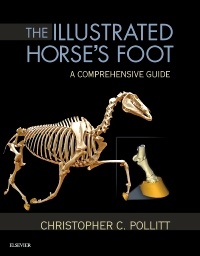
Subject for The Illustrated Horse's Foot:

Publication date: 12-2015
272 p. · 21.4x27.6 cm · Hardback
272 p. · 21.4x27.6 cm · Hardback
Description
/li>Contents
/li>
Achieve optimal results in equine foot care and treatment! The Illustrated Horse's Foot: A Comprehensive Guide uses clear instructions in an atlas-style format to help you accurately identify, diagnose, and treat foot problems in horses. Full-color clinical photographs show structure and function as well as the principles of correct clinical examination and shoeing, and a companion website has videos depicting equine foot cases. Written by internationally renowned expert Christoher Pollitt, this resource enhances your ability to treat equine conditions ranging from laminitis to foot cracks, infections, trauma, vascular compromise, and arthritis.
- Comprehensive coverage addresses a wide range of equine foot conditions.
- A unique collection of MIMICs provides beautifully detailed anatomical hoof images.
- 284 high-quality images show conditions of the equine foot, including many 2-D reconstructions of MRI and CT data.
- Step-by-step case histories follow equine patients from initial presentation through diagnosis to treatment and outcome.
- A convenient, templated format provides quick access to clinical signs, diagnosis, treatment, and prognosis.
- Expert author Chris Pollitt is a pioneer in the use of advanced radiographic, CT, and MRI technology for imaging equine foot and laminitis problems to facilitate accurate diagnosis and effective treatment.
- A companion website located at pollitthorsesfoot.com located at pollitthorsesfoot.com includes video clips of equine foot cases.
SECTION I: Foot Structure and Function
1. The Hoof
2. The Hoof Capsule
3. The Dermis
4. The MIMICS Anatomical Models
5. Planar Anatomy
6. The Suspensory Apparatus of the Distal Phalanx
7. The Circulatory System
8. Histology
9. Electron Microscopy
10. Bones
11. Joints
12. Radiography of the Foot
13. The Palmar Digital Nerve
SECTION II: Conditions of the Foot
14. Laminitis
15. Navicular Disease
16. Midline Toe Cracks
17. Seedy Toe ('White Line Disease')
18. Ossification of the Ungular Cartilages
19. Coronary Band Injury
20. Infected Nail Holes
© 2024 LAVOISIER S.A.S.
These books may interest you
Equine Internal Medicine 241.27 €