Imaging from Cells to Animals In Vivo Series in Cellular and Clinical Imaging Series
Coordonnateurs : Barroso Margarida, Intes Xavier
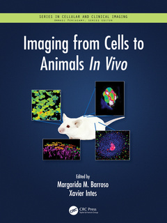
Imaging from Cells to Animals In Vivo offers an overview of optical imaging techniques developed over the past two decades to investigate biological processes in live cells and tissues. It comprehensively covers the main imaging approaches used as well as the application of those techniques to biological investigations in preclinical models. Among the areas covered are cell metabolism, receptor-ligand interactions, membrane trafficking, cell signaling, cell migration, cell adhesion, cytoskeleton and other processes using various molecular optical imaging techniques in living organisms, such as mice and zebrafish.
Features
- Brings together biology and advanced optical imaging techniques to provide an overview of progress and modern methods from microscopy to whole body imaging.
- Fills the need for a comprehensive view of application-driven development and use of new tools to ask new biological questions in the context of a living system.
- Includes basic chapters on key methods and instrumentation, from fluorescence microscopy and imaging to endoscopy, optical coherence tomography and super-resolution imaging.
- Discusses approaches at different length scales and biomedical applications to the study of single cell, whole organ, and whole organism behavior.
- Addresses the impact on discovery, such as cellular function as implicated in human disease and translational medicine, for example in cancer diagnosis.
Section I: Overview of Imaging Methods and Instrumentation. 1. Fluorescence microscopy techniques. 2. Intra-vital microscopy, M Perro. 3. An introduction to live-cell super-resolution imaging. 4. Endoscopic Optical Coherence Tomography: Technologies and Applications. 5. Bioluminescence. 6. Macroscopic Fluorescence Imaging. 7. Optical Coherence Tomography. 8. Multiscale Photoacoustic Imaging. 9. Fluorescence Lifetime: Techniques, Analysis & Applications in the Life Sciences. Section II: Imaging cellular behavior. 10. Imaging cell metabolism. 11. Intravital imaging of cancer cell migration in vivo. 12. Imaging cellular signaling in vivo using fluorescent protein biosensors. 13. Imaging cell adhesion and migration. 14. Imaging the living eye. Section III: Whole-organ and whole-organism imaging. 15. Heart imaging. 16. Visualizing hepatic immunity through the eyes of intravital microscopy. 17. Optical imaging of the mammalian oviduct in vivo. 18. Immune system imaging. 19. Brain imaging in live mice. 20. Live imaging of zebrafish. 21. Whole-body fluorescence imaging in cancer research. 22. Large-scale fluorescence imaging in neuroscience
Margarida Barroso is a Professor at the Department of Molecular and Cellular Physiology, Albany Medical College (Albany, NY).
Xavier Intes is a Professor in the Biomedical Engineering Department and Co-Director of the Center for Modeling, Simulation and Imaging for Medicine (CeMSIM),at Rensselaer Polytechnic Institute (Troy, U.S.).
Date de parution : 05-2023
21x28 cm
Date de parution : 12-2020
21x28 cm
Thèmes d’Imaging from Cells to Animals In Vivo :
Mots-clés :
OCT Image; Fluorescence Lifetimes; biomedical imaging; Multiphoton Microscopy; fluorescence lifetime microscopy; Intravital Microscopy; Optical imaging techniques; Vivo Imaging; Transgenic animals; Optical Coherence Tomography; Biological processes; FLIM; Fluorescence microscopy; Axial Resolution; Live cells; Intravital Imaging; Vivo OCT Imaging; OCT System; Light Sheet; Sim; Zebrafish Embryo; Pam; CTC; LSM; OCT Endoscope; Fluorescence Imaging; Structural OCT Image; Photon Counting; PAI; Sted Microscopy; FLIM Imaging; Photoacoustic Signals