


Description
Cone Beam Computed Tomography
Imaging in Medical Diagnosis and Therapy Series
Coordinator: Shaw Chris C.
Language: English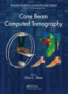
Subjects for Cone Beam Computed Tomography:
Keywords
Image Guided Radiation Therapy; Digital Breast Tomosynthesis; Image Visualization; CBCT; Image Analysis; CBCT System; Image Processing; CBCT Image; Image Simulation; Bow Tie Filter; Image Correction; Cone Beam; Small Animal Imaging; Proc SPIE; Radiation Treatment; Projection Image; Interventional Radiology; Cone Beam Reconstruction; Perception; Radiation Dose; CT System; Artifacts; X-ray Tube; Image Acquisition; Tube Current; Reconstruction; Tube Current Modulation; Image Quality; CBCT Data; Imaging; Antiscatter Grid; X-Ray; CBCT Scan; Medical; Glandular Dose; CT; Cardiac Phase; Cone Beam CT; Scatter X-rays; Helical Scan; Computed Tomography; Axial Scan; Gantry Rotation Time; MONTE CARLO

Publication date: 02-2014
Support: Print on demand
Publication date: 06-2020
· 21x28 cm · Paperback
Description
/li>Contents
/li>Biography
/li>
Conventional computed tomography (CT) techniques employ a narrow array of x-ray detectors and a fan-shaped x-ray beam to rotate around the patient to produce images of thin sections of the patient. Large sections of the body are covered by moving the patient into the rotating x-ray detector and x-ray source gantry. Cone beam CT is an alternative technique using a large area detector and cone-shaped x-ray beam to produce 3D images of a thick section of the body with one full angle (360 degree or 180 degree plus detector coverage) rotation. It finds applications in situations where bulky, conventional CT systems would interfere with clinical procedures or cannot be integrated with the primary treatments or imaging systems.
Cone Beam Computed Tomography explores the past, present, and future state of medical x-ray imaging while explaining how cone beam CT, with its superior spatial resolution and compact configuration, is used in clinical applications and animal research. The book:
- Supplies a detailed introduction to cone beam CT, covering basic principles and applications as well as advanced techniques
- Explores state-of-the-art research and future developments while examining the fundamental limitations of the technology
- Addresses issues related to implementation and system characteristics, including image quality, artifacts, radiation dose, and perception
- Reviews the historical development of medical x-ray imaging, from conventional CT techniques to volumetric 3D imaging
- Discusses the major components of cone beam CT: image acquisition, reconstruction, processing, and display
A reference work for scientists, engineers, students, and imaging professionals, Cone Beam Computed Tomography provides a solid understanding of the theory and implementation of this revolutionary technology.
Part I: Fundamental Principles and Techniques. History of X-Ray Computed Tomography: From Fan-Beam to Cone Beam. Acquisition of Projection Images. Reconstruction Algorithms. Cone Beam CT Image Quality. Radiation Dose. Part II: Advanced Techniques. Image Simulation. 3D Image Processing, Analysis, and Visualization. Volume-of-Interest Cone Beam CT. 4D Cone Beam CT. Image Correction. Part III: Applications. Multi-Detector Row CT. Cone Beam Micro-CT for Small Animal Research. Cardiac Imaging. C-Arm CT in the Interventional Suite. Cone Beam CT: Transforming Radiation Treatment Guidance, Planning, and Monitoring. Breast CT.
Chris C. Shaw is a professor and the director of digital imaging research at the University of Texas M. D. Anderson Cancer Center, Houston. He received his Ph.D from the University of Wisconsin-Madison under the supervision of Charles A. Mistretta. As a graduate student, he participated in Dr. Mistretta’s research in developing the digital subtraction angiography (DSA) technique. He has since been working on the development and investigation of new medical x-ray imaging techniques, including cone beam CT and digital tomosynthesis, for applications to angiography, mammography, chest imaging, and interventional imaging.
These books may interest you
Statistics of Medical Imaging 232.80 €