


Description
Cyto- and Myeloarchitectural Brain Atlas of the Ferret (Mustela putorius) in MRI Aided Stereotaxic Coordinates, 1st ed. 2018
Author: Radtke-Schuller Susanne
Language: English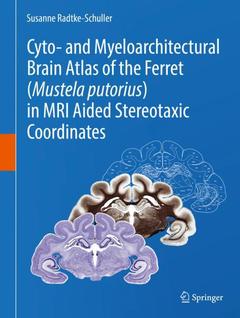
Subjects for Cyto- and Myeloarchitectural Brain Atlas of the Ferret...:
Keywords
Neuroanatomy; Carnivore; Cytoarchitecture; Fiber architecture; MRI
242.64 €
In Print (Delivery period: 15 days).
Add to cart the print on demand of Radtke-Schuller Susanne
Support: Print on demand
Description
/li>Contents
/li>Biography
/li>Comment
/li>
Description
This stereotaxic atlas of the ferret brain provides detailed architectonic subdivisions of the cortical and subcortical areas in the ferret brain using high-quality histological material stained for cells and myelin together with in vivo magnetic resonance (MR) images of the same animal. The skull-related position of the ferret brain was established according to in vivo MRI and additional CT measurements of the skull. Functional denotations from published physiology and connectivity studies are mapped onto the atlas sections and onto the brain surface, together with the architectonic subdivisions. High-resolution MR images are provided at levels of the corresponding histology atlas plates with labels of the respective brain structures. The book is the first atlas of the ferret brain and the most detailed brain atlas of a carnivore available to date. It provides a common reference base to collect and compare data from any kind of research in the ferret brain.
Key Features
- Provides the first ferret brain atlas with detailed delineations of cortical and subcortical areas in frontal plane.
- Provides the most detailed brain atlas of a carnivore to date.
- Presents a stereotaxic atlas coordinate system derived from high-quality histological material and in vivo magnetic resonance (MR) images of the same animal.
- Covers the ferret brain from forebrain to spinal cord at intervals of 0.6 mm on 58 anterior-posterior levels with 5 plates each.
- Presents cell (Nissl) stained frontal sections (plate 1) and myelin stained sections (plate 2) in a stereotaxic frame.
- Provides detailed delineations of brain structures and their denomination on a Nissl stained background on a separate plate (3).
- Compiles abbreviations on plate 4, a plate that also displays the low resolution MRI of the atlas brain with the outlines of the Nissl sections in overlay.
- Displays high-resolution MR images at intervals of 0.15 mm from another animal with labeled brain structures as plate 5 corresponding to the anterior-posterior level of each atlas plate.
- Provides detailed references used for delineation of brain areas.
Target audience of the book:
The book addresses researchers and students in neurosciences who are interested in brain anatomy in general (e.g., for translational purposes/comparative aspects), particularly those who study the ferret as important animal model of growing interest in neurosciences.
Introduction.- Methods.- Index of Brain Structures.- Surface Views of the Ferret Brain.- Atlas Plates with Sub-Panels.
A leading neuroanatomist long affiliated with the University of Munich (LMU München), now with the University of North Carolina at Chapel Hill (UNC), Dr. Radtke-Schuller has developed a new approach to brain atlases. She recently received high praise from one of Germany’s leading neuroanatomists, Prof. Karl Zilles (Jülich), for her gerbil brain atlas, published as a supplement to the journal Brain Structure & Function.
She has worked in cooperation with well-known international scientists on the neuroanatomy and physiology of bats and tenrecs and, for more than a decade, on ferret neuroanatomy.
First ever cyto- and myeloarchitectural brain atlas of the ferret (Mustela putorius)
New approach combines high resolution histology images with MRI in a common stereotaxic reference coordinate system
Resulting reference coordinates provide a highly accurate anatomical database as a foundation for research
Important animal model for neuroscience research
These books may interest you
Stereotaxic and Chemoarchitectural Atlas of the Brain of the Common Marmoset (Callithrix jacchus) 220.72 €
Neuroanatomy text and atlas 69.87 €