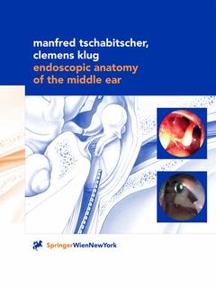
Description
Endoscopic Anatomy of the Middle Ear, Softcover reprint of the original 1st ed. 2000
Authors: Tschabitscher Manfred, Klug Clemens
Language: English
Subjects for Endoscopic Anatomy of the Middle Ear:
Approximative price 158.24 €
In Print (Delivery period: 15 days).
Add to cart
Endoscopic Anatomy of the Middle Ear
Publication date: 11-2012
130 p. · 21x27.9 cm · Paperback
Publication date: 11-2012
130 p. · 21x27.9 cm · Paperback
Approximative price 158.24 €
Subject to availability at the publisher.
Add to cart
Endoscopic anatomy of the middle ear
Publication date: 09-2000
130 p. · 21x27.9 cm · Hardback
Publication date: 09-2000
130 p. · 21x27.9 cm · Hardback
Description
/li>Contents
/li>Comment
/li>
Filling a gap in the anatomical and ENT literature, the authors show the various approaches to the middle ear which allow safe surgical manipulations, such as through the tympanic membrane or the Eustachian tube.
A Transmeatal approach (4 mm rigid scope).- A 1.1 Site of approach.- A 1.2 Following the chorda tympani (70° scope).- A 1.3 View into the anterior epitympanic recess (30°, 70° scopes).- A 1.4 Overview of protympanum (30° scope).- A 1.5 Central mesotympanum seen through 70°, 30°, 0° scopes.- A 1.6 Incus (30°, 70° scopes).- A 1.7 Stapes (70° scope).- A 1.8 Mucosal folds surrounding ossicular chain (70° scope).- A 1.9 Posterior wall of tympanic cavity (70° scope).- A 1.10 Epitympanic recess (70° scope).- A 1.11 Hypotympanum (30° scope).- A 1.12 Some variants of round window niche (30°, 70° scopes).- B Transtympanic approach.- B 1 Anterior inferior transtympanic approach.- B 1.1 Site of approach.- B 1.2 Overview of protympanum from the top down (rigid scope 2.7 mm, 30°).- B 1.3.1 Protympanic region close to tympanic membrane (rigid scope, 1.9 mm, 30°).- B 1.3.2 Medial wall of protympanum and tympanic opening of EUSTACHIAN tube (rigid scope, 1.9 mm, 30°).- B 1.4.1 Center of protympanum (rigid scope, 1.9 mm, 60°).- B 1.4.2 Roof of protympanum (rigid scope, 1.9 mm, 60°).- B 1.4.3 Bottom of protympanum (rigid scope, 1.9 mm, 60°).- B 2 Posterior superior transtympanic approach (1.9 mm rigid scope).- B 2.1 Site of approach.- B 2.2.1 Course of chorda tympani.- B 2.2.2 Posterior wall with pyramidal eminence (30° scope).- B 2.2.3 Facial canal (30° scope).- B 2.3.1 Oval window niche (30° scope).- B 2.3.2 Stapes (30° scope).- B 2.3.3 Anterior tympanic isthmus (30° scope).- B 2.4 Anterior epitympanic recess (60° scope).- B 4.5 Aditus ad antrum (60° scope).- B 2.6 Protympanum and hypotympanum (60° scope).- B 3 Posterior inferior transtympanic approach (1.9 mm rigid scope).- B 3.1 Site of approach.- B 3.2.1 Approaching the round window niche (30° scope).- B 3.2.2 Secondary tympanic membrane (30° scope).- B 3.3.1 Stapes ( 30° scope).- B 3.3.2 View into vestibule (stapes removed) (30° scope).- B 3.4 Posterior wall of tympanic cavity (30° scope).- B 3.5 Posterior wall (60° scope).- B 3.6 Stapes seen from below (60° scope).- B 3.7 Roof and medial wall of protympanum (60° scope).- B 3.8 Floor of protympanum and hypotympanum (60° scope).- C Transmastoid approach.- C 1 Through antrum.- C 1.1 Site of approach.- C 1.2 Antrum mastoideum (rigid scope, 4 mm, 0°).- C 1.3 View into mesotympanum from the top down (rigid scope, 2.7 mm, 30°).- C 1.4.1 Incudostapedial joint and anterior tympanic isthmus (rigid scope, 1.9 mm, 30°).- C 1.4.2 Cochleariform process and anterior epitympanic recess (rigid scope, 1.9 mm, 30°).- C 1.5.1 Incudomallear joint and chorda tympani (rigid scope, 1.9 mm, 60°).- C 1.5.2 Stapes and posterior tympanic isthmus (rigid scope, 1.9 mm, 60°).- C 2 Through facial recess (1.9 mm rigid scope).- C 2.1 Site of approach.- C 2.2.1 Tympanic membrane (30° scope).- C 2.2.2 Pyramidal eminence (30° scope).- C 2.2.3 Facial canal (30° scope).- C 2.2.4 Promontory and round window niche (30° scope).- C 2.2.5 Hypotympanum.- C 2.3.1 Round window niche (60° scope).- C 2.3.2 Tympanic ring and tympanic membrane (60° scope).- C 2.3.3 PRUSSAK’s space (60° scope).- D Transtubal approach (0.8 mm flexible steerable scope).- D 1.1 Site of approach.- D 1.2.1 View from tympanic opening of EUSTACHIAN tube into mesotympanum.- D 1.2.2 Scope kept straight to approach mesotympanic structures.- D 1.2.3 Scope advanced into central mesotympanum.- D 1.2.4 Scope crosses over stapes (medially to incus) and looks into mesotympanum.- D 1.2.5 Scope between long limb of incus and facial canal: Aditus ad antrum.- D 1.3.1 Scope in mesotympanum bent laterally: Lateral wall of tympanic cavity.- D 1.3.2 Scope in mesotympanum bent laterally and upward: PRUSSAK’s space.- D 1.3.3 Scope in mesotympanum bent laterally and downward: Hypotympanum.- D 1.4.1 Scope in protympanum bent upward: Mucosal fold of tensor tympani.- D 1.4.2 Scope advanced: Perforation of mucosal fold of tensor tympani.- D 1.4.3 Scope above tendon of tensor tympani: Incudomallear joint.- D 1.4.4 Scope medial to incudomallear joint: Posterior epitympanic recess.- D 1.5.1 Scope below tendon of tensor tympani: Incudostapedial joint.- D 1.5.2 Scope medial to long limb of incus: Facial canal and lateral semicircular canal.- D 1.5.3 Scope crosses over stapes: Aditus ad antrum.- D 1.6.1 Scope bent laterally and upward behind handle of malleus: Chorda tympani.- D 1.6.2 Scope lateral to long limb of incus: Course of chorda tympani.- D 1.7.1 Scope in the plane of the handle of malleus: Stapes.- D 1.7.2 Scope below incudostapedial joint: Posterior wall.- E Two-port approach (1.9 mm rigid scope, 30° ’ 0.8 mm flexible steerable scope).- E 1.1 Transtubal scope crosses above the tendon of the tensor tympani.- E 1.2 Transtubal scope crosses below the tendon of the tensor tympani.- E 1.3 Transtubal scope looks at the ossicular chain.- E 1.4 Transtubal scope looks at the posterior wall.
Color Atlas (200 coloured figures)
No endoscopic anatomy available
Fills a gap in the anatomical and ENT literature
© 2024 LAVOISIER S.A.S.


