Description
Essentials of Clinical Anatomy of the Equine Locomotor System
Author: Denoix Jean-Marie
Language: English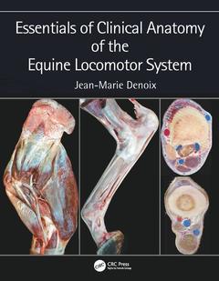
Subjects for Essentials of Clinical Anatomy of the Equine Locomotor...:
Keywords
Sesamoidean Ligament; Digital Flexor Tendon; horse; Superficial Digital Flexor Tendon; cross-sectional anatomy; Deep Digital Flexor Tendon; regional and topographical anatomy; Medial Collateral Ligament; clinical anatomy; Lateral Digital Extensor Tendon; diagnostic imaging; ultrasonography; MRI images and CT scans; Digital Extensor Tendon; dissections; Proximal Sesamoid Bone; Locomotor system; Oblique Sesamoidean Ligament; Diagnostic imaging; Fourth Metacarpal Bone; Distal thoracic limb; Accessory Carpal Bone; Equine locomotor system; Common Calcanean Tendon; Extensor Carpi Radialis Muscle; Interosseous Muscle; Collateral Ligament; Distal Digital Annular Ligament; Distal Sesamoid Bone; Dorsal Digital Extensor Tendon; Digital Annular Ligament; Central Tarsal Bone; Collateral Sesamoidean Ligament; Extensor Carpi Radialis Tendon; Palmar Eminence; Fourth Metatarsal Bone; Caudal Ramus

· 17.8x25.4 cm · Hardback
Description
/li>Contents
/li>Readership
/li>Biography
/li>
Essentials ofClinical Anatomy of the Equine Locomotor System presents a unique photographic record of dissections showing the topographical anatomy of the locomotor system of the horse. Readers of this book will be able to see the position and relationships of the bones, joints, muscles, nerves and blood vessels that make up each region of the forelimb, vertebral column and hindlimb.
Key features:
- Important features of regional and topographical anatomy are presented using full-color photos of detailed dissections
- Anatomy is presented in a clinical context
- Preparations of cross-sectional anatomy facilitate interpretation of diagnostic imaging, such as ultrasonography, MRI images and CT scans
- All dissections are of fresh material, rather than preserved specimens, to demonstrate the appearance of tissues in the living animal, or at post mortem autopsy
This new atlas is essential for anybody involved in detailed anatomical study, complex lameness evaluation or advanced imaging techniques in horses. It will be a useful guide for veterinary students, and a reference for equine vets in practice.
Preface. Chapter 1. FORELIMB: Foot, Pastern, Fetlock, Metacarpus, Carpus, Forearm, Elbow, Arm, Shoulder. Chapter 2. VERTEBRAL COLUMN: Neck, Back, Pelvis. Chapter 3. HINDLIMB: Digital area, Fetlock, Metatarsus, Tarsus, Crus, Stifle, Thigh, Hip. Index.
Jean-Marie Denoix graduated from the Veterinary School of Lyon (France, 1977). Agrégé in Veterinary Anatomy (1983), he defended a PhD thesis in Biomechanics (1987). In 1988 he moved to the Veterinary School of Alfort and became professor of Anatomy, head of the equine clinical unit (until 2005) and head of a research unit devoted to Biomechanics and Pathology of the locomotor system in horses (until 2003). He became head of CIRALE (Center of Imaging and Research in Equine Locomotor Pathology in Horses) in 1999. Since 2006 he is President of the ISELP (International Society of Equine Locomotor Pathology), large animal imaging associate of the ECVDI since 2010, and President of the ALAPILE (South American Association) since 2012. In 2013 he became Diplomate of the American College of Veterinary Sport Medicine and Rehabilitation (ACVSMR) and is a founding Diplomate of the European College of Veterinary Sport Medicine and Rehabilitation (ECVSMR).