Description
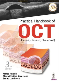
Practical Handbook of OCT (3rd Ed.)
(Retina, Choroid, Glaucoma)
Authors: Rispoli Marco, Savastano Maria Cristina, Lumbroso Bruno
Language: English
Subjects for Practical Handbook of OCT:
121.50 €
In Print (Delivery period: 14 days).
Add to cart204 p. · 21.5x27.8 cm · Paperback
Description
/li>Contents
/li>Biography
/li>
Optical coherence tomography (OCT) is a non-invasive imaging test that uses light waves to take cross-sectional pictures of the retina, the light-sensitive tissue lining the back of the eye (eyeSmart). The technique is recognised worldwide as an essential device for diagnosis, assessment and follow up of retinal diseases and glaucoma.
The third edition of this comprehensive manual has been fully revised to provide clinicians and trainees with the most recent advances in OCT imaging. New examination and diagnostic protocols are covered in depth and this edition includes a step by step guide to data interpretation.
Divided into three sections, the book begins with discussion on interpretation of OCT images, including ?en face? and dyeless angiography.
The second section covers lesions and diseases, and part three explains new syndromes and classifications.
Highly illustrated with clinical images and tables, this practical reference has been written by renowned experts based in Italy.
Key points
- Practical guide to recent advances in OCT imaging
- Fully revised, new edition covers new examination and diagnostic protocols, with step by step guide to data interpretation
- Internationally recognised, Italy-based author team
- Previous edition (9789351525318) published in 2014
Section 1: Methods of OCT interpretation
- Chapter 1: Practical suggestions to obtain clear and clinically useful optical coherence tomography images
- Chapter 2: Basic normal anatomy and optical coherence tomography
- Chapter 3: Logical method of optical coherence tomography interpretation: analysis and synthesis
- Chapter 4: Tridimensional and ‘en face’ scan analysis
- Chapter 5: Optical coherence tomography dyeless angiography
- Chapter 6: Synthesis and deduction
Section 2: Elementary lesions and frequent diseases
- Chapter 7: Elementary optical coherence tomography lesions
- Chapter 8: Ocular syndromes and disorders: more frequent disorders
- Chapter 9: Ocular syndromes and disorders: less frequent disorders
- Chapter 10: Complex case analysis and interpretation
- Chapter 11: Glaucoma
- Chapter 12: Neurodegenerative diseases and optical coherence tomography
Section 3: New syndromes, new classifications
- Chapter 13: Contribution of optical coherence tomography to new syndromes description and new clinical classifications
- Chapter 14: Closing comments: the future
Suggested reading
Index
Marco Rispoli MD
Surgery and Emergency Unit, Rome Eye Hospital, Centro Italiano Macula, Rome, Italy
Maria Cristina Savastano MD PhD
Clinical Research Coordinator, Ophthalmology Unit, Policlinico Universitario A Gemelli, Catholic University, Rome, Centro Italiano Macula, Rome, Italy
Bruno Lumbroso MD
Centro Italiano Macula, Rome, Italy
These books may interest you

Practical Retinal OCT 74.00 €


