Description
Retina Atlas, New edition
Authors: Grewal SPS, Brar Manpreet, Sharma Mansi, Dogra Mangat R, Grewal Dilraj S
Language: English
Subjects for Retina Atlas:
290 p. · 24.1x33 cm · Hardback
Out of Print
Description
/li>Contents
/li>Biography
/li>
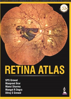
This atlas provides ophthalmologists with a collection of images to help with the identification, diagnosis and subsequent treatment of retinal disorders.
The images are procured from Eidon scanner technology and also include optical coherence tomography (OCT) pictures to assist with understanding of related pathologies.
Divided into nine sections, the book begins with images illustrating the normal fundus. Each of the following sections covers a different retinal disorder including diabetic retinopathy, macula disorders, retinal detachment, ocular tumours and hereditary diseases.
Each section features a multitude of images, each with brief descriptive text to assist understanding.
Key points
- Comprehensive atlas of retinal imaging for diagnosis of ocular disorders
- Images procured from Eidon scanner technology
- Includes OCT images to assist understanding of related pathologies
- Covers many different retinal disorders and diseases
1. Normal Fundus
2. Diabetic Retinopathy
3. Retinal Vascular Disorders
4. Macula
5. Retinal Detachment
6. Inflammatory
7. Hereditary
8. Ocular Tumours and Optic Nerve Disorders
9. Miscellaneous
SPS Grewal MBBS MD
CEO, Grewal Eye Institute, Chandigarh, India
Manpreet Brar MBBS MS
Senior Consultant, Department of Vitreo-retinal Diseases and Surgery, Grewal Eye Institute, Chandigarh, India
Mansi Sharma MBBS DNB FAICO
Consultant, Department of Vitreo-retinal Diseases and Surgery, Grewal Eye Institute, Chandigarh, India
Mangat R Dogra MBBS MS
Director, Department of Vitreo-retinal Diseases and Surgery, Grewal Eye Institute, Chandigarh, India
Dilraj S Grewal MBBS MD
Associate Professor of Ophthalmology, Vitreoretinal Surgery and Uveitis, Duke Eye Centre, Duke University Medical Centre, Durham, North Carolina, USA


