


Description
Morphological Mouse Phenotyping
Anatomy, Histology and Imaging
Authors: Ruberte Jesus, Carretero Ana, Navarro Marc
Language: English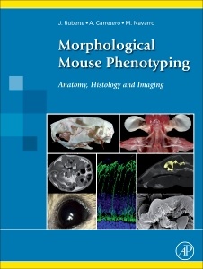
Subjects for Morphological Mouse Phenotyping:
Keywords
<; P>; Mouse morphology; mouse anatomy; mouse atlas; mouse histology; mouse imaging<; /P>
600 p. · 30.4x22.8 cm · Hardback
Out of Print
Description
/li>Contents
/li>Readership
/li>Biography
/li>Comment
/li>
Morphological Mouse Phenotyping: Anatomy, Histology and Imaging is an atlas of explanatory diagrams and text that guides the reader through normal mouse anatomy, histology, and imaging. The book is targeted for mouse researchers and veterinarian and human pathologists, and presents a complete, integrative description of normal mouse morphology.
Disease animal models are fundamental in research to improve human health. The success of using genetically engineered mice to evaluate molecular disease hypotheses has encouraged the development of massive global projects, making the mouse the most used animal disease model. Laboratory mouse populations are straining the housing capacity of pharmaceutical and biotechnology companies, as well as public research institutions. However, the scientific community lacks sufficient expertise in morphological phenotyping to effectively characterize and validate these animal models. The mouse displays fundamental morphological similarities to humans; however, a mouse is not a man.
1. Introduction 2. Osteology 3. Arthrology 4. Myology 5. Digestive tract 6. Respiratory apparatus 7. Urinary organs 8. Male genital organs 9. Female genital organs 10. Anatomy of development 11. Circulatory System 12. The endocrine glands 13. Nervous System 14. Eye and related structures 15. Vestibulocochlear orga 16. Common integument
scientists working with mice; laboratory animal vets.
Ana Carretero works with the Mouse Imaging Platform in the Center for Animal Biotechnology and Gene Therapy, Department of Animal Health and Anatomy, Veterinary School, Universitat Autònoma de Barcelona, Spain.
- Features more than 2,200 original images showing the anatomy, histology, and cellular structure of mouse organs
- Includes images specifically produced for this book in the Mouse Imaging Platform (Center for Animal Biotechnology and Gene Therapy, Universitat Autònoma de Barcelona)
- Offers an integrative vision of mouse morphology using correlative X-ray, computed tomography, magnetic resonance, and ultrasound images
- Employs classical anatomical techniques such as conventional dissection, skeletal preparations, vascular injections, and histological, immunohistochemical, and electron microscopy techniques to characterize mouse morphology
These books may interest you

The Laboratory Mouse 71.13 €