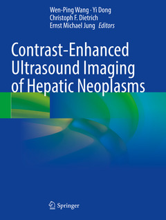
Contrast-Enhanced Ultrasound Imaging of Hepatic Neoplasms, 1st ed. 2021
Langue : Anglais
Coordonnateurs : Wang Wen-Ping, Dong Yi, Dietrich Christoph F., Jung Ernst Michael

This book aims to provide reader an overview of clinical applications of contrast-enhanced ultrasound in hepatic neoplasms diagnosis. Ultrasound images and pathological results of different hepatic neoplasms are introduced in the chapters, including benign liver tumors, malignant liver tumors, hepatic carcinoma, intrahepatic cholangiocarcinoma, rare liver benign and malignant neoplasms, regenerative nodules, inflammatory pseudotumor, parasite liver lesions, and hepatitis peliosis, etc. The combination of ultrasound findings with final histopathological results then discover the potential mechanical of contrast enhancement changes.
With the development of ultrasound technology and widely application of ultrasound contrast agents (USCA) in recent decades, contrast-specific imaging modalities have been developed in combination with USCA and a low mechanical index (MI), allowing continuous real-time grey scale imaging. The updated contrast-specific software for liver diseases and hepatic tumors diagnosis has also been described described in detail. With high-resolution contrast ultrasound images during arterial phase, portal venous phase and late phase, author wants to show the whole dynamic wash-in and wash-out process of the different focal liver lesions.
This book is an invaluable resource for radiologists, hepatologists and oncologists in their everyday clinical practice.
Liver Contrast Enhanced Ultrasound-How to Perform it.- Benign Liver Tumors.- Malignant Liver Tumors.- Rare Benign Liver Tumors.- Rare Malignant Liver Tumors.- Regenerative Nodules, Low Grade and High Grade Dysplastic Nodules in Liver Cirrhosis.- Parasite Liver Lesions.- Inflammatory Psedotumour.- Hepatitis Peliosis.- Dynamic Vascular Pattern and Quantitative Analysis in Liver Tumors.- Application of Volume Navigation and Imaging Fusion in Liver Tumors.- 3D-CEUS Liver Tumors.- CEUS and Interventional Treatment of Liver Tumors.- Future Directions and Challenges.
Wen-Ping Wang is a Professor and Chief at the Department of Ultrasound, Zhongshan Hospital affiliated to Fudan University, Shanghai, China.
Yi Dong is a Professor at the same department with Wen-Ping Wang.
Christoph F. Dietrich is a Professor at Department Allgemeine Innere Medizin (DAIM), Kliniken Hirslanden Beau Site, Salem und Permancence, Bern, Switzerland.
Ernst-Michael Jung is a Professor at Institut für Röntgendiagnostik, Universitätsklinikum Regensburg, Regensburg, Germany.
Includes contrast-enhanced ultrasound images and pathological results of different hepatic neoplasms Accompanies specialist comments in each case A useful reference for radiologists and hepatologists
Date de parution : 06-2022
Ouvrage de 269 p.
21x27.9 cm
Date de parution : 06-2021
Ouvrage de 269 p.
21x27.9 cm
Disponible chez l'éditeur (délai d'approvisionnement : 15 jours).
Prix indicatif 189,89 €
Ajouter au panierThème de Contrast-Enhanced Ultrasound Imaging of Hepatic Neoplasms :
© 2024 LAVOISIER S.A.S.


