Description
CT Colonography Atlas, Softcover reprint of the original 1st ed. 2013
For the Practicing Radiologist
Diagnostic Imaging Series
Coordinators: Neri Emanuele, Faggioni Lorenzo, Bartolozzi Carlo
Language: English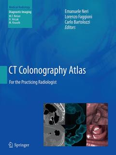
Subjects for CT Colonography Atlas:
Keywords
CT Colonography; Cancer; Colon; Colonoscopy; Image Processing

Publication date: 08-2016
Support: Print on demand
Publication date: 07-2013
184 p. · 21x27.9 cm · Hardback
Description
/li>Contents
/li>Comment
/li>
This easy-to-use atlas comprises a collection of representative common and unusual virtual colonoscopy (CT colonography, CTC) cases that physicians and radiologists may expect to encounter during their clinical practice. The atlas reflects the important recent advances in image acquisition, patient preparation, and image processing and is thus completely up-to-date. Each case is presented with the native CT images, integrated images obtained by 3D image processing, and colonoscopic correlation. Topics covered include normal appearances, anatomical variants, pitfalls, diverticula, lipomas, inflammatory bowel disease, polyps, flat lesions, cancers, and the postsurgical colon. By presenting the main features of anatomy and pathology, this atlas will serve as an invaluable tool both for radiologists performing CTC and for clinicians who need to review the CTC examinations of their patients.
Normal Colon.- Anatomical Variants.- Pitfalls.- Diverticula.- Lypoma.-Inflammatory Bowel Diseases.- Polyps: Pedunculated.- Polyps: Sessile.- Flat Lesions.- Colon Cancer.- Rectal Cancer.- Cancers of the Ileo-Cecal Valve.- Post-Surgical Colon.
These books may interest you
Atlas of Virtual Colonoscopy 116.04 €