Description
Cytohistology of Focal Liver Lesions
Author: Wee Aileen
Language: English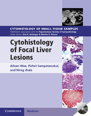
Approximative price 183.09 €
In Print (Delivery period: 14 days).
Add to cart the book of Wee Aileen
Publication date: 03-2014
Description
/li>Contents
/li>
Each volume in this richly illustrated series, published in association with the Papanicolaou Society of Cytopathology, provides an organ-based approach to the cytologic and histologic diagnosis of small tissue samples. Benign, pre-malignant and malignant entities are presented in a well-organized and standardized format, with high-resolution color photomicrographs, tables, and lists of key specific morphologic criteria. Example vignettes allow the reader to assimilate the diagnostic principles in a case-based format. This volume provides comprehensive coverage of both surgical pathology and cytopathology of focal liver lesions. Extensively illustrated throughout, it contains key cytologic and histologic features, practical points, radiologic and morphologic pictures, flow charts, and tabulated summaries for easy comprehensive overview and quick reference and provides a pragmatic algorithmic approach to cytohistologic diagnosis. With over 700 printed photomicrographs and a CD-ROM offering all images in a downloadable format, this is an important resource for all anatomic pathologists.
Preface; 1. The focal liver lesion: general considerations; 2. Morphologic approach; 3. Diagnostic algorithm; 4. Focal liver lesions with low or no suspicion of malignancy; 5. Focal liver lesions suspicious of hepatocellular carcinoma; 6. Focal liver lesions suspicious of intrahepatic cholangiocarcinoma; 7. Focal liver lesions from metastases and other malignancies; 8. Focal liver lesions with cystic appearance; 9. Focal liver lesions in infants and children; 10. Ancillary studies; 11. Techniques and technology in practice; Index.
© 2024 LAVOISIER S.A.S.
These books may interest you
Pancreatic Cytohistology 161.07 €