Description
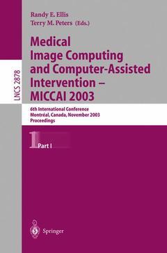
Medical Image Computing and Computer-Assisted Intervention - MICCAI 2003, 2003
6th International Conference, Montréal, Canada, November 15-18, 2003, Proceedings, Part I
Lecture Notes in Computer Science Series, Vol. 2878
Coordinators: Ellis Randy E., Peters Terry M.
Language: English
Subjects for Medical Image Computing and Computer-Assisted...:
Publication date: 10-2003
822 p. · 15.5x23.5 cm · Paperback
822 p. · 15.5x23.5 cm · Paperback
Description
/li>Contents
/li>Comment
/li>
The 6th International Conference on Medical Imaging and Computer-Assisted Intervention,MICCAI2003,washeldinMontr? eal,Qu? ebec,CanadaattheF- rmont Queen Elizabeth Hotel during November 15?18, 2003. This was the ?rst time the conference had been held in Canada. The proposal to host MICCAI 2003 originated from discussions within the Ontario Consortium for Ima- guided Therapy and Surgery, a multi-institutional research consortium that was supported by the Government of Ontario through the Ontario Ministry of E- erprise, Opportunity and Innovation. The objective of the conference was to o?er clinicians and scientists a - rum within which to exchange ideas in this exciting and rapidly growing ?eld. MICCAI 2003 encompassed the state of the art in computer-assisted interv- tions, medical robotics, and medical-image processing, attracting experts from numerous multidisciplinary professions that included clinicians and surgeons, computer scientists, medical physicists, and mechanical, electrical and biome- cal engineers. The quality and quantity of submitted papers were most impressive. For MICCAI 2003 we received a record 499 full submissions and 100 short c- munications. All full submissions, of 8 pages each, were reviewed by up to 5 reviewers, and the 2-page contributions were assessed by a small subcomm- tee of the Scienti?c Review Committee. All reviews were then considered by the MICCAI 2003 Program Committee, resulting in the acceptance of 206 full papers and 25 short communications. The normal mode of presentation at MICCAI 2003 was as a poster; in addition, 49 papers were chosen for oral presentation.
Simulation and Planning.- The Role of Simulation Fidelity in Laparoscopic Surgical Training.- Simulation Studies for Predicting Surgical Outcomes in Breast Reconstructive Surgery.- Atlas-Based Recognition of Anatomical Structures and Landmarks to Support the Virtual Three-Dimensional Planning of Hip Operations.- Pathology Growth Model Based on Particles.- Needle Steering and Model-Based Trajectory Planning.- Brain Shift Correction Based on a Boundary Element Biomechanical Model with Different Material Properties.- Mesh Topology Identification for Mass-Spring Models.- A New Biomechanical Model Based Approach on Brain Shift Compensation.- Real-Time Synthesis of Bleeding for Virtual Hysteroscopy.- A Biomechanical Model of the Liver for Reality-Based Haptic Feedback.- Image-Based Modelling of Soft Tissue Deformation.- Individualized Geometric Model from Unorganized 3-D Points: An Application to Thorax Modeling.- Highly Accurate CAD Tools for Cranial Implants.- Medially Based Meshing with Finite Element Analysis of Prostate Deformation.- An “Optimal” k-Needle Placement Strategy Given an Approximate Initial Needle Position.- Robotic Mechanism ans Mechanical Properties of Tissue.- Automatic Targeting Method and Accuracy Study in Robot Assisted Needle Procedures.- A New Haptic Sensor Actuator System for Virtual Reality Applications in Medicine.- Simple Biomanipulation Tasks with “Steady Hand” Cooperative Manipulator.- A Transurethral Prostate Resection Manipulator for Minimal Damage to Mucous Membrane.- Virtual Remote Center of Motion Control for Needle Placement Robots.- Optimum Robot Control for 3D Virtual Fixture in Constrained ENT Surgery.- Interactive Guidance by Image Overlay in Robot Assisted Coronary Artery Bypass.- Comparison of Registration Procedures of the Tibia in Robot-Assisted Total Knee Arthroplasty.- A New Method to Extend Applicable Area of Minimally Invasive Neurosurgery by Brain Retract Manipulator.- Evaluating the Role of Vision and Force Feedback in Minimally Invasive Surgery: New Automated Laparoscopic Grasper and A Case Study.- Characterization of Intra-abdominal Tissues from in vivo Animal Experiments for Surgical Simulation.- Measurement-Based Deep Venous Thrombosis Screening System.- Determination of the Mechanical Properties of Soft Human Tissues through Aspiration Experiments.- Episode Classification for the Analysis of Tissue/Instrument Interaction with Multiple Visual Cues.- In-Vivo and Postmortem Compressive Properties of Porcine Abdominal Organs.- Application of an Intra-operative Load Measuring System for Knee Replacement Surgery.- Modelling and Optimization of Bone-Cutting Forces in Orthopaedic Surgery.- Soft Tissue Simulation Based on Measured Data.- Analysis of Forces during Robotic Needle Insertion to Human Vertebra.- A Modular 2-DOF Force-Sensing Instrument For Laparoscopic Surgery.- Interventional Registration.- Intensity-Based 2D-3D Spine Image Registration Incorporating One Fiducial Marker.- Application of XMR 2D-3D Registration to Cardiac Interventional Guidance.- 3D Elastic Registration of Vessel Lumen from IVUS Data on Biplane Angiography.- pq-Space Based 2D/3D Registration for Endoscope Tracking.- Accuracy of a Fluoroscopy Technique for Assessing Patellar Tracking.- Design and Implementation of Parallel Nonrigid Image Registration Using Off-the-Shelf Supercomputers.- Vascular Atlas Formation Using a Vessel-to-Image Affine Registration Method.- The Creation of a Brain Atlas for Image Guided Neurosurgery Using Serial Histological Data.- Effective Intensity-Based 2D/3D Rigid Registration between Fluoroscopic X-Ray and CT.- A Spatial-Stiffness Analysis of Fiducial Registration Accuracy.- Temporal Lobe Epilepsy Lateralization Based on MR Image Intensity and Registration Features.- Model-Updated Image Guidance: A Statistical Approach to Gravity-Induced Brain Shift.- Registration of Organ Surface with Intra-operative 3D Ultrasound Image Using Genetic Algorithm.- Exploring RSA Ultimate Accuracy by Using Computer Synthetic Images.- New Image Similarity Measure for Bronchoscope Tracking Based on Image Registration.- Diffusion Tensor and Functional MRI Fusion with Anatomical MRI for Image-Guided Neurosurgery.- Cardiac Imaging.- 4-D Tomographic Representation of Coronary Arteries from One Rotational X-Ray Sequence.- Flow Field Abstraction and Vortex Detection for MR Velocity Mapping.- Automated Segmentation of the Left Ventricle in Cardiac MRI.- Segmentation of 4D Cardiac MR Images Using a Probabilistic Atlas and the EM Algorithm.- ICA vs. PCA Active Appearance Models: Application to Cardiac MR Segmentation.- Four-Chamber 3-D Statistical Shape Model from Cardiac Short-Axis and Long-Axis MR Images.- Tracking Atria and Ventricles Simultaneously from Cardiac Short- and Long-Axis MR Images.- Exploratory Identification of Cardiac Noise in fMRI Images.- Optic Flow Computation from Cardiac MR Tagging Using a Multiscale Differential Method.- A Finite Element Model for Functional Analysis of 4D Cardiac-Tagged MR Images.- Cardiac Endoscopy Enhanced by Dynamic Organ Modeling for Minimally-Invasive Surgery Guidance.- Automated Model-Based Segmentation of the Left and Right Ventricles in Tagged Cardiac MRI.- Algorithms for Real-Time FastHARP Cardiac Function Analysis.- Automatic Segmentation of Cardiac MRI.- Cardiac LV Segmentation Using a 3D Active Shape Model Driven by Fuzzy Inference.- Automatic Planning of the Acquisition of Cardiac MR Images.- A High Resolution Dynamic Heart Model Based on Averaged MRI Data.- Analysis of Left Ventricular Motion Using a General Robust Point Matching Algorithm.- Segmentation I.- Interactive, GPU-Based Level Sets for 3D Segmentation.- 3D Image Segmentation of Deformable Objects with Shape-Appearance Joint Prior Models.- A Novel Stochastic Combination of 3D Texture Features for Automated Segmentation of Prostatic Adenocarcinoma from High Resolution MRI.- An Automatic System for Classification of Nuclear Sclerosis from Slit-Lamp Photographs.- Multi-scale Nodule Detection in Chest Radiographs.- Automated White Matter Lesion Segmentation by Voxel Probability Estimation.- Drusen Detection in a Retinal Image Using Multi-level Analysis.- 3D Automated Lung Nodule Segmentation in HRCT.- Segmentation and Evaluation of Adipose Tissue from Whole Body MRI Scans.- Automatic Identification and Localization of Craniofacial Landmarks Using Multi Layer Neural Network.- An Artificially Evolved Vision System for Segmenting Skin Lesion Images.- Multivariate Statistics for Detection of MS Activity in Serial Multimodal MR Images.- Vascular Attributes and Malignant Brain Tumors.- Statistical-Based Approach for Extracting 3D Blood Vessels from TOF-MyRA Data.- Automated Segmentation of 3D US Prostate Images Using Statistical Texture-Based Matching Method.- Clinical Applications of Medical-Image Computing.- An Evaluation of Deformation-Based Morphometry Applied to the Developing Human Brain and Detection of Volumetric Changes Associated with Preterm Birth.- Statistical Shape Modeling of Unfolded Retinotopic Maps for a Visual Areas Probabilistic Atlas.- Optimal Scan Planning with Statistical Shape Modelling of the Levator Ani.- Determining Epicardial Surface Motion Using Elastic Registration: Towards Virtual Reality Guidance of Minimally Invasive Cardiac Interventions.- A CAD System for Quantifying COPD Based on 3-D CT Images.- Temporal Subtraction of Thorax CR Images.- Computer Aided Diagnosis for CT Colonography via Slope Density Functions.- Disease-Oriented Evaluation of Dual-Bootstrap Retinal Image Registration.- The Navigated Image Viewer – Evaluation in Maxillofacial Surgery.- Lung Deformation Estimation with Non-rigid Registration for Radiotherapy Treatment.- Registration, Matching, and Data Fusion in 2D/3D Medical Imaging: Application to DSA and MRA.- Texture Analysis of MR Images of Minocycline Treated MS Patients.- Estimating Cortical Surface Motion Using Stereopsis for Brain Deformation Models.- Automatic Spinal Deformity Detection Based on Neural Network.
Includes supplementary material: sn.pub/extras
© 2024 LAVOISIER S.A.S.


