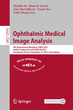
Ophthalmic Medical Image Analysis, 1st ed. 2021
8th International Workshop, OMIA 2021, Held in Conjunction with MICCAI 2021, Strasbourg, France, September 27, 2021, Proceedings

Image Processing, Computer Vision, Pattern Recognition, and Graphics Series

Coordinators: Fu Huazhu, Garvin Mona K., MacGillivray Tom, Xu Yanwu, Zheng Yalin

Language: English
Approximative price 68.56 €

In Print (Delivery period: 15 days).

Add to cartAdd to cart
Publication date:
200 p. · 15.5x23.5 cm · Paperback

This book constitutes the refereed proceedings of the 8th International Workshop on Ophthalmic Medical Image Analysis, OMIA 2021, held in conjunction with the 24th International Conference on Medical Imaging and Computer-Assisted Intervention, MICCAI 2021, in Strasbourg, France, in September 2021.*

The 20 papers presented at OMIA 2021 were carefully reviewed and selected from 31 submissions. The papers cover various topics in the field of ophthalmic medical image analysis and challenges in terms of reliability and validation, number and type of conditions considered, multi-modal analysis (e.g., fundus, optical coherence tomography, scanning laser ophthalmoscopy), novel imaging technologies, and the effective transfer of advanced computer vision and machine learning technologies.

*The workshop was held virtually.


Adjacent Scale Fusion and Corneal Position Embedding for Corneal Ulcer Segmentation.- Longitudinal detection of diabetic retinopathy early severity grade changes using deep learning.- Intra-operative OCT (iOCT) Image Quality Enhancement: A Super-Resolution Approach using High Quality iOCT 3D Scans.- Diabetic Retinopathy Detection based on Weakly Supervised Object Localization and Knowledge Driven Attribute Mining.- FARGO: A Joint Framework for FAZ and RV Segmentation from OCTA Images.- CDLRS: Collaborative Deep Learning Model with Joint Regression and Segmentation for Automatic Fovea Localization.- U-Net with Hierarchical Bottleneck Attention for Landmark Detection in Fundus Images of the Degenerated Retina.- Radial U-Net: Improving DMEK Graft Detachment Segmentation in Radial AS-OCT Scans.- Guided Adversarial Adaptation Network for Retinal and Choroidal Layer Segmentation.- Juvenile Refractive Power Prediction based on Corneal Curvature and Axial Length via a Domain Knowledge Embedding Network.- Peripapillary Atrophy Segmentation with Boundary Guidance.- Are cardiovascular risk scores from genome and retinal image complementary? A deep learning investigation in a diabetic cohort.- Dual-branch Attention Network and Atrous Spatial Pyramid Pooling for Diabetic Retinopathy Classification Using Ultra-Widefield Images.- Self-Adaptive Transfer Learning for Multicenter Glaucoma Classification in Fundus Retina Images.- Multi-Modality Images Analysis: A Baseline for Glaucoma Grading via Deep Learning.- Impact of data augmentation on retinal OCT image segmentation for diabetic macular edema analysis.- Representation and Reconstruction of Image-Based Structural Patterns of Glaucomatous Defects Using Only Two Latent Variables from a Variational Autoencoder.- Stacking Ensemble Learning in Deep Domain Adaptation for Ophthalmic Image Classification.- Attention Guided Slit Lamp Image Quality Assessment.- Robust Retinal Vessel Segmentation from a Data Augmentation Perspective.