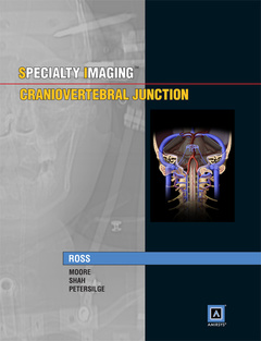
Specialty Imaging: Craniovertebral Junction is a unique reference tool for a unique topic! The craniovertebral junction is imaged on every brain, neck and cervical spine MR and CT study performed. This region has unique nomenclature, embryology, anatomy, vasculature, biomechanics and pathology. Surgical techniques are also used in this region that are distinctive, and related to the underlying complex anatomy.
Specialty Imaging: Craniovertebral Junction incorporates normal anatomy, embryology, specialized imaging techniques and the myriad of unique pathology which occurs in that region
. with the time-saving bulleted text and state-of-the art annotated radiographic and medical illustrations, this volume will surely prove to be an indispensable resource for residents and fellows in radiology, neurosurgery, and orthopaedic surgery who deal with the skull base and craniocervical / craniovertebral junction. In addition, Amirsys eBook Advantage™ supplements the printed edition by providing an additional imaging dimension to each diagnosis, fully searchable text, and the ease of electronic access.
FEATURES:
§ Numerous full color medical illustrations, state-of-the-art imaging, and pathologic correlation
§ Features the classic benefits of all Amirsys® titles, including time-saving bulleted text, Key Facts in each chapter, stunning annotated images, and an extensive index
§ Amirsys eBook Advantage™, an online version of the print book with hundreds of additional images, larger images, and fully searchable text


