Description
Coherent Raman Scattering Microscopy
Series in Cellular and Clinical Imaging Series
Coordinators: Cheng Ji-Xin, Xie Xiaoliang Sunney
Language: English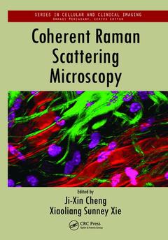
Subjects for Coherent Raman Scattering Microscopy:
Keywords
Ch Ap; Car Microscopy; Coherent Raman Scattering; Car Imaging; Label-Free Imaging; Car Signal; Point-By-Point Chemical Map Of Live Cells And Tissues; Stokes Beams; Crs Microscopy; Fatty Acyl CoA; Nonlinear Optical Spectroscopy; Non-resonant Background; Resonant Signals; Ampli Er; Multiplex Car; Crs; Neutral Lipid Droplets; Spontaneous Raman Spectrum; CH2 Symmetric Stretch Vibration; Nonresonant Signal; Local Oscillator; Car Spectrum; Pump Beam; Probe Pulses; Lipid Droplets; Raman Resonance; SHG Microscopy; Coherent anti-Stokes Raman Spectroscopy; Cytoplasmic Lipid Droplets; Spontaneous Raman Scattering

Publication date: 04-2018
· 17.8x25.4 cm · Paperback
Publication date: 11-2012
550 p. · 17.8x25.4 cm · Hardback
Description
/li>Contents
/li>Readership
/li>Biography
/li>
The First Book on CRS Microscopy
Compared to conventional Raman microscopy, coherent Raman scattering (CRS) allows label-free imaging of living cells and tissues at video rate by enhancing the weak Raman signal through nonlinear excitation. Edited by pioneers in the field and with contributions from a distinguished team of experts, Coherent Raman Scattering Microscopy explains how CRS can be used to obtain a point-by-point chemical map of live cells and tissues.
In color throughout, the book starts by establishing the foundation of CRS microscopy. It discusses the principles of nonlinear optical spectroscopy, particularly coherent Raman spectroscopy, and presents the theories of contrast mechanisms pertinent to CRS microscopy. The text then provides important technical aspects of CRS microscopy, including microscope construction, detection schemes, and data analyses. It concludes with a survey of applications that demonstrate how CRS microscopy has become a valuable tool in biomedicine.
Due to its label-free, noninvasive examinations of living cells and organisms, CRS microscopy has opened up exciting prospects in biology and medicine?from the mapping of 3D distributions of small drug molecules to identifying tumors in tissues. An in-depth exploration of the theories, technology, and applications, this book shows how CRS microscopy has impacted human health and will deepen our understanding of life processes in the future.
Theory: Theory of Coherent Raman Scattering. Coherent Raman Scattering under Tightly Focused Conditions. Platforms: Construction of a Coherent Raman Microscope. Stimulated Raman Scattering Microscopy. Femtosecond versus Picosecond Pulses for Coherent Raman Microscopy. Wide-Field CARS Microscopy. Vibrational Spectromicroscopy by Coupling Coherent Raman Imaging with Spontaneous Raman Spectral Analysis. Coherent Control in CARS. Fourier Transform CARS Microscopy. CRS with Alternative Beam Profiles. Vibrational Phase Microscopy. Multiplex CARS Microscopy. Interferometric Multiplex CARS. Photonic Crystal Fiber-Based Broadband CARS Microscopy. Multiplex Stimulated Raman Scattering Microscopy. Applications: Imaging Myelin Sheath Ex Vivo and In Vivo by CARS Microscopy. Imaging Lipid Metabolism in Caenorhabditis elegans and Other Model Organisms. Lipid-Droplet Biology and Obesity-Related Health Risks. White Matter Injury: Cellular-Level Myelin Damage Quantification in Live Animals. CARS Microscopy Study of Liquid Crystals. Live Cell Imaging by Multiplex CARS Microspectroscopy. Coherent Raman Scattering Imaging of Drug Delivery Systems. Applications of Stimulated Raman Scattering Microscopy. Applications of Coherent Anti-Stokes Raman Spectroscopy Imaging to Cardiovascular Diseases. Applications of CARS Microscopy to Tissue Engineering. Dietary Fat Absorption Visualized by CARS Microscopy. Index.
Ji-Xin Cheng is a professor of biomedical engineering at Purdue University. He earned a PhD from the University of Science and Technology of China and completed postdoctoral research at the Hong Kong University of Science and Technology and Harvard University. He is a pioneer in the development and biomedical application of molecular vibration-based imaging tools.
Xiaoliang Sunney Xie is a Mallinckrodt Professor of Chemistry at Harvard University. He earned a PhD from the University of California-San Diego and completed postdoctoral research at the University of Chicago. He is well-known for his innovations in nonlinear Raman microscopy and his pioneering work in single-molecule biophysical chemistry, including enzyme dynamics and live cell gene expression.