Description
Atlas of Clinical Imaging and Anatomy of the Equine Head
Authors: Kimberlin Larry, zur Linden Alex, Ruoff Lynn
Language: English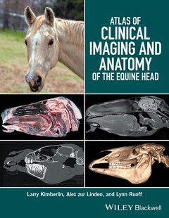
Subject for Atlas of Clinical Imaging and Anatomy of the Equine Head:
153.93 €
In Print (Delivery period: 14 days).
Add to cart the book of Kimberlin Larry, zur Linden Alex, Ruoff Lynn
160 p. · 21.3x27.9 cm · Hardback
Description
/li>Contents
/li>Biography
/li>
- Provides a comprehensive comparative atlas to structures of the equine head
- Pairs gross anatomy with radiographs, CT, and MRI images
- Presents an image-based reference for understanding anatomy and pathology
- Covers radiography, computed tomography, and magnetic resonance imaging
Introduction: General Presentation of Atlas, vi
1 Overview of CT and MRI of the Equine Head, 1
2 Clinical and Surgical Anatomy of the Equine Head: Transverse Sections, 9
3 Clinical and Surgical Anatomy of the Equine Head: Sagittal Sections, 85
Brain sagittal close-up, 100
4 Clinical and Surgical Anatomy of the Equine Head: Dorsal Sections, 115
Glossary, 143
References, 145
Index, 147
Larry Kimberlin, DVM, FAVD, CVPP, is the owner of Northeast Texas Veterinary Dental Center in Greenville, Texas, USA.
Alex zur Linden, DVM, DACVR, is Assistant Professor of Radiology at Ontario Veterinary College at the University of Guelph in Ontario, Canada.
Lynn Ruoff, DVM is Clinical Associate Professor at Texas A&M University’s College of Veterinary Medicine in College Station, Texas, USA.
These books may interest you
Color Atlas of Equine Pathology 164.63 €