Description
Clinical Radiology of the Horse (4th Ed.)
Authors: Butler Janet A., Colles Christopher M., Dyson Sue J., Kold Svend E., Poulos Paul W.
Language: English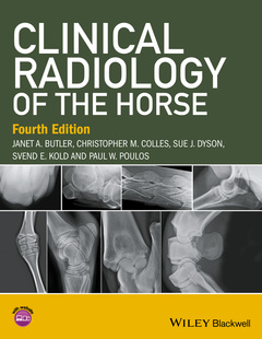
Subject for Clinical Radiology of the Horse:
Keywords
equine x-ray
226.23 €
In Print (Delivery period: 14 days).
Add to cart the book of Butler Janet A., Colles Christopher M., Dyson Sue J., Kold Svend E., Poulos Paul W.
816 p. · 21.8x27.9 cm · Hardback
Description
/li>Contents
/li>Biography
/li>
Clinical Radiology of the Horse is the best-selling, practical guide to all areas of equine radiography and radiology written by an experienced group of clinicians with a broad range of backgrounds.
- Offers an atlas of normal and clinical images, as well as a comprehensive guide to techniques, equipment, positioning, and interpretation for general veterinary practitioners and specialists in imaging and orthopaedics
- Updates to this fourth edition fully reflect the move to digital imaging with many new figures in the book and major revisions to the chapters on the head, thorax, and abdomen
- Contains expanded coverage of the foot, pastern, and fetlock (now in separate chapters)
- Includes a password-protected website with all the images from the book as well as over 200 additional images with examples of more subtle lesions, more fractures, correct technique and positioning versus incorrect, immature horses, progression of disease, and pathological images
About the authors v
Preface to the fourth edition vii
About the companion website ix
1 General principles 1
Introduction, 1
Principles of radiography, 3
Principles of radiographic interpretation: radiology 15
Radiological appearance of physiological changes and some common pathological lesions, 20
2 Computed and digita l radiography 41
3 The foot_55
Distal phalanx (pedal bone), 55
Hoof, 97
Navicular bone, 113
4 The proximal and middle phalanges and the proximal interphalangeal joint_149
5 Metacarpopha langeal and metatarsopha langeal (fet lock) joints_175
6 The metacarpal and metatarsal regions_215
7 The carpus and antebrach ium_259
8 The shoulder, humerus , elbow and radius_301
Scapulohumeral (shoulder) joint and humerus, 301
Humeroradial,humeroulnar and radioulnar (elbow or cubital) joints and radius, 330
9 The tarsus_349
10 The stifle and tibia_399
Stifle, 399
Tibia, 440
11 The head 449
Cranium, 451
Paranasal sinuses (frontal, maxillary, conchal) and maxilla, 466
Teeth and mandible, 480; Pharynx, larynx and Eustachian tube diverticulum, 512
12 The vertebra l column 531
Cervical vertebrae, 531
Thoracolumbar vertebrae, 569
Sacrum and coccygeal vertebrae, 602
13 The pelvis and femur_609
Pelvis, 609
Femur, 632
14 The thorax 639
15 The alimentar y and urinary systems_687
Oesophagus, 694; Abdomen and gastrointestinal tract, 705
Urinary system, 715
16 Miscellaneous techniques_723
Arthrography and bursography, 723
Tendonography, 724
Angiography, 725;
Venography, 730
Myelography, 733
Pneumocystography, 743
Intravenous Pyelography, 744
Other techniques, 744
Appendix A: Fusion times of physes and suture lines_749
Appendix B: Exposure guide, image quality and film processing faults_753
Appendix C: Glossary 761
Index 767
Janet A. Butler
Jan specialises in equine radiography and has 40 years’ experience in this field. She joined the Animal Health Trust in Newmarket, UK in 1975 where she gained considerable experience working with many internationally renowned veterinary surgeons. Since 1997 she has been working in private practice, initially at the Willesley Equine Clinic, UK, which since 2009 has been part of the B&W Equine Group.
Christopher M. Colles
Chris qualified from the Royal Veterinary College, UK in 1971. After three years in mixed practice (where he obtained a Part I Diploma in Radiology) he joined the Animal Health Trust as a clinician in 1975. He has carried out research in many areas of equine orthopaedics and radiology, having a particular interest in the horse’s foot. In 1988 he returned to practice, where he became a senior partner in Avonvale Veterinary Practice, specialising in equine orthopaedics, until his recent retirement from practice. He is recognised by the Royal College of Veterinary Surgeons as a Specialist in Equine Orthopaedic Surgery. Chris was awarded an Honorary Fellowship of the Worshipful Company of Farriers in 2000 in recognition of his research into conditions of the foot, and involvement with farriery education.
Sue J. Dyson
After qualifying from the University of Cambridge in 1980, Sue worked for a year at New Bolton Center, University of Pennsylvania, and then spent a year in private practice in Pennsylvania. Sue then joined the Centre for Equine Studies of the Animal Health Trust, UK, where she has specialised in lameness diagnosis and diagnostic imaging. Sue is recognised as a Specialist in Equine Orthopaedics by the Royal College of Veterinary Surgeons and holds the RCVS Diploma in Equine Orthopaedics. She is an Associate of the European College of Veterinary Diagnostic Imaging. She has published widely on lameness, radiography ultrasonography, nuclear scintigraphy and magnetic resonan