Description
Micro-Tomographic Atlas of the Mouse Skeleton, Softcover reprint of the original 1st ed. 2007
Authors: Bab Itai A., Hajbi-Yonissi Carmit, Gabet Yankel, Müller Ralph
Language: English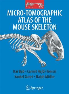
Subjects for Micro-Tomographic Atlas of the Mouse Skeleton:
Keywords
Atlas; Bab; Mouse; Müller; Skeleton; Tempo; Tomographic; anatomy; bone; computed tomography (CT)
Approximative price 210.99 €
In Print (Delivery period: 15 days).
Add to cart the print on demand of Bab Itai A., Hajbi-Yonissi Carmit, Gabet Yankel, Müller Ralph
Publication date: 08-2016
Support: Print on demand
Approximative price 210.99 €
Subject to availability at the publisher.
Add to cart the book of Bab Itai A., Hajbi-Yonissi Carmit, Gabet Yankel, Müller Ralph
Publication date: 09-2007
205 p. · 21x27.9 cm · Hardback
Description
/li>Contents
/li>Comment
/li>
The Micro-Tomographic Atlas of the Mouse Skeleton provides a unique systematic description of all calcified components of the mouse. It includes about 200 high resolution, two and three dimensional m CT images of the exterior and interiors of all bones and joints. In addition, the spatial relationship of bones within complex skeletal units is also described. The images are accompanied by detailed explanatory text, thus highlighting special features and newly reported structures. The Atlas fulfils an emerging need for a comprehensive reference to assist both trained and in-training researchers.
Detailed images accompanied by explanatory text, which describe the microstructure of the mouse skeleton.
So far a systematic two- and tri-dimensional descriptions of the internal anatomy of bones, as well as the tri-dimensional relationship exhibited in joints, is not available.
These books may interest you
MRI Atlas of Normal Anatomy 52.74 €
Atlas of the Developing Mouse Brain 146.54 €