Description
Atlas of the Developing Mouse Brain (2nd Ed.)
Authors: Paxinos George, Halliday Glenda, Watson Charles, Kassem Mustafa S.
Language: English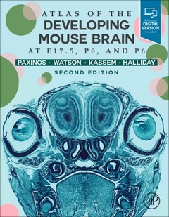
Subjects for Atlas of the Developing Mouse Brain:
146.54 €
In Print (Delivery period: 14 days).
Add to cart the book of Paxinos George, Halliday Glenda, Watson Charles, Kassem Mustafa S.
Publication date: 06-2020
384 p. · 21.4x27.6 cm · Hardback
384 p. · 21.4x27.6 cm · Hardback
Description
/li>Contents
/li>Readership
/li>Biography
/li>Comment
/li>
Atlas of the Developing Mouse Brain, Second Edition builds on the features of successful first edition, providing a comprehensive and convenient reference for all areas of the mouse brain at Fetal-Day 17.5 (E17.5), Day-of-Birth (P0), and Day-Six postnatal (P6). The book also delineates the parts of the eye, features of the skull, ganglia, nerves, arteries, veins, bones and foramina. This atlas is an essential tool for researchers and students who study the development of the mouse brain, or for those who interpret findings from genetic manipulation.
Preface to the second editionReproduction of altas figures in other publicationsAcknowledgementsIntroductionHistologyPreparation of photographs and drawingsThe construction of abbreviations in the Paxinos/Watson nomenclatureIdentification of structuresReferencesList of structuresIndex of abbreviationsFigures
Standard neuroanatomy lab atlas for all labs doing neuroanatomy, neurophysiology, neuropharmacology, behavioral neuroscience, or molecular expression/cloning/neurogenetics in the mouse brain
Professor Paxinos is the author of almost 50 books on the structure of the brain of humans and experimental animals, including The Rat Brain in Stereotaxic Coordinates, now in its 7th Edition, which is ranked by Thomson ISI as one of the 50 most cited items in the Web of Science. Dr. Paxinos paved the way for future neuroscience research by being the first to produce a three-dimensional (stereotaxic) framework for placement of electrodes and injections in the brain of experimental animals, which is now used as an international standard. He was a member of the first International Consortium for Brain Mapping, a UCLA based consortium that received the top ranking and was funded by the NIMH led Human Brain Project. Dr. Paxinos has been honored with more than nine distinguished awards throughout his years of research, including: The Warner Brown Memorial Prize (University of California at Berkeley, 1968), The Walter Burfitt Prize (1992), The Award for Excellence in Publishing in Medical Science (Assoc Amer Publishers, 1999), The Ramaciotti Medal for Excellence in Biomedical Research (2001), The Alexander von Humbolt Foundation Prize (Germany 2004), and more
Glenda Halliday works at the University of Sydney in NSW, Sydney, Australia..
Charles Watson is a neuroscientist and public health physician. His qualifications included a medical degree (MBBS) and two research doctorates (MD and DSc). He is Professor Emeritus at Curtin University, and holds adjunct professorial research positions at the University of New South Wales, the University of Queensland, and the University of Western Australia.
He has published over 100 refereed journal articles and 40 book chapters, and has co-authored over 25 books on brain and spinal cord anatomy. The Paxinos Watson rat brain atlas has been cited over 80,000 times. His current research is focused on the comparative anatomy of the hippocampus and the claustrum.
He was awarded the degree of Doctor of Science by the University
- Contains 176 high-resolution color scans of Nissl-stained coronal sections of the brain and skull of the fetal (E17.5), day-of-birth (P0), and day-six postnatal mouse (P6)
- Includes diagrams that delineate all structures of the brain, as well as peripheral nerves, ganglia, muscles, bones, veins and arteries of the head
- Presents approximately 5000 corrections and updates from the first edition
- Includes color codes of the veins, arteries, nerves and ganglions of the skull in diagrams
© 2024 LAVOISIER S.A.S.
These books may interest you
Netter's Atlas of Neuroscience 69.56 €