Description
Cross-Sectional Atlas of the Human Head, 1st ed. 2017
With 0.1-mm pixel size color images
Author: Park Jin Seo
Language: English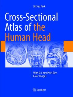
Subjects for Cross-Sectional Atlas of the Human Head:

Publication date: 06-2019
328 p. · 21x27.9 cm · Paperback
Publication date: 01-2018
Support: Print on demand
Description
/li>Contents
/li>Biography
/li>Comment
/li>
This superb color atlas sets a new standard in neuroanatomy by presenting around 300 detailed thin-sectioned images of the human head, including the brain, with 0.1-mm intervals and a pixel size of 0.1 mm × 0.1 mm. A new reference system employed for this purpose is clearly explained, and structures are fully annotated in the horizontal, coronal, and sagittal planes. Recent advances in 7T MRI and 7T TDI have considerably enhanced imaging of the human brain, thereby impacting on both neuroscience research and clinical practice. Moreover, the information gained from initiatives involving photography of thin slices of human cadavers, such as the Visible Human Projects, Visible Korean and Chinese Visible Human, has enriched knowledge of neuroanatomy and thereby facilitated the interpretation of such ultra-high-field resolution images. The exquisite images contained within this atlas will be invaluable in providing both researchers and clinicians with important new insights.
Introduction.- Horizontal plane.- Coronal plane.- Sagittal plane.- Index.