Description
The Equine Distal Limb (2nd Ed.)
An Atlas of Clinical Anatomy and Comparative Imaging
Author: Denoix Jean-Marie
Language: English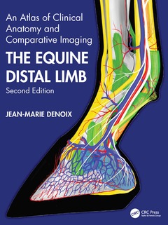
Subjects for The Equine Distal Limb:
Keywords
sesamoidean; ligament; deep; digital; flexor; tendon; superficial; dorsal; ramus; annular

· 21x28 cm · Hardback
Description
/li>Contents
/li>Readership
/li>Biography
/li>
Jean-Marie Denoix is the world?s leading equine musculoskesletal system anatomist and has become one of the foremost equine diagnostic ultrasonographers. There is therefore nobody better to compile a reference atlas of the clinical anatomy of the foot, pastern and fetlock, correlated with images obtained by radiography, diagnostic ultrasonography and magnetic resonance imaging. Advanced imaging techniques require in depth knowledge of anatomy for accurate interpretation and especially when using magnetic resonance imaging this must be a 3-dimensional concept of anatomy.
This new edition replaces existing ultrasound images and most of the radiographic and MRI images with new, updated versions and adds new images of extraordinarily high quality, the multiple views of each area of the distal limb providing an extremely detailed evaluation. Each part opens with a characteristically superb anatomical drawing by the author. Each spread of pages deals with a single dissection viewed by means of colour photographs, labelled black and white equivalents, plus x-rays, ultrasound and MRI scans as required.
The diagnosis and management of distal limb lameness require a precise knowledge of the functional anatomy and biomechanics of the equine distal joints, ligaments and tendons which are presented in the last chapter. The book is designed for maximum clarity using a generous page size. The atlas is essential for anybody involved in detailed anatomical study, complex lameness evaluation or advanced imaging techniques.
Foreword. Preface. Acknowledgements. General Presentation of the Atlas. Overview of the Distal Limb Anatomy 1. The Equine Foot. Dissections of the Equine Foot. Sagittal and Parasagittal Sections of the Equine Foot. Transverse Sections of the Equine Foot. Frontal Sections of the Equine Foot. The Equine Pastern. Dissections of the Equine Pastern. Sagittal and Parasagittal Sections of the Equine Pastern. Transverse Sections of the Equine Pastern. Frontal Sections of the Equine Pastern. 2. The Equine Fetlock. Dissections of the Equine Fetlock. Sagittal and Parasagittal Sections of the Equine Fetlock. Transverse Sections of the Equine Fetlock. Frontal Sections of the Equine Fetlock. Functional Anatomy of the Equine Distal Limb. Glossary of English and Latin Equivalents. Index.
Jean-Marie DENOIX Professor, DVM, PhD, Founder ISELP, DACVSMR, DECVSMR.
For more than forty years, Pr J-M Denoix has been captivated by equine musculoskeletal system anatomy and biomechanics and was involved in the diagnosis and rehabilitation of equine lameness. He become assistant of Anatomy with Professor R. Barone in 1978 at the National Veterinary School of Lyons where he presented his PhD research on the biomechanics of the equine distal limb in 1987. He moved to the National Veterinary School of Alfort in 1988 to become head of the Anatomy and Equine departments. Founder of CIRALE (Center of Imaging and Research on Equine Locomotor Affections) in Normandy, France in 1999, he founded ISELP (International Society of Equine Locomotor Pathology) in 2006 and ALAPILE (Latinoamerican Association of Pathology and Imaging of the locomotor system in Equine) in 2012. In 2013 he became Diplomate of the American College of Veterinary Sport Medicine and Rehabilitation (ACVSMR) and is a founding Diplomate of the European College of Veterinary Sport Medicine and Rehabilitation (ECVSMR). In 2018, he set up a rehabilitation unit at CIRALE where residents of the American and European Colleges of Veterinary Sport Medicine and Rehabilitation are trained.
Jean-Marie has been an invited speaker at many international meetings in more than 30 countries around the world. In 2021, as a recognition of his career and achievements in the field of biomechanics and lameness in horses, he has been invited to present the ‘John Hickman Plenary lecture’ at the BEVA meeting and the State of the Art lecture (‘Milne Lecture’) at the AAEP convention.